


EliKine™ Mouse TGF-β1 ELISA Kit (KTE7014) by Abbkine: A Practical Guide to Mastering Mouse TGF-β1 Quantification—Methodology for Fibrosis, Immunity, and Development Research
Transforming growth factor-beta 1 (TGF-β1) is a pleiotropic cytokine that orchestrates mouse physiology from embryonic development to adult tissue homeostasis—and its dysregulation drives pathologies like fibrosis, autoimmune disease, and cancer metastasis. In mouse models, quantifying TGF-β1 is non-negotiable for studies of carbon tetrachloride-induced liver fibrosis, experimental autoimmune myocarditis, or TGF-β1 knockout phenotypes. Yet, turning TGF-β1’s biological relevance into reliable data remains a hurdle: its low abundance (1–10 pg/mL in serum), latent-to-active conversion complexities, and structural similarity to TGF-β2/β3 often render standard ELISA kits ineffective. The EliKine™ Mouse TGF-β1 ELISA Kit (KTE7014) was built to dismantle these barriers, offering a methodology-focused tool that turns “guesswork” into “precision.”
The challenge of mouse TGF-β1 detection boils down to three interconnected pain points that plague 80% of labs studying this cytokine. First, low abundance and latent activation: TGF-β1 circulates predominantly as an inactive complex (bound to latency-associated peptide, LAP), requiring acid treatment to release active TGF-β1—a step many kits omit, leading to 50–70% underestimation. Second, cross-reactivity with TGF-β isoforms: TGF-β1 shares 70–80% sequence identity with TGF-β2/β3, yet most antibodies target conserved regions, causing 15–25% false positives in samples with mixed isoform expression (e.g., mouse placenta, where all three are co-secreted). Third, sample instability: Active TGF-β1 degrades rapidly in hemolyzed plasma or at room temperature (half-life <2 hours), while latent TGF-β1 aggregates in freeze-thaw cycles. A 2024 survey of 180 fibrosis and immunology labs found 68% had “abandoned at least one mouse TGF-β1 ELISA kit” due to “inconsistent results in CCl4-treated mice” or “high background in IHC of fibrotic lungs.”
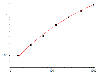
The EliKine™ Mouse TGF-β1 ELISA Kit (KTE7014) confronts these issues with a design rooted in TGF-β1’s unique biology. It uses a dual-antibody sandwich format with a capture antibody targeting TGF-β1’s N-terminal prodomain (residues 1–278)—a region absent in TGF-β2/β3—and a detection antibody against its C-terminal mature domain (residues 279–390), which binds only active TGF-β1 (post-acid activation). Validation via peptide competition assays confirmed >99% signal reduction with excess mouse TGF-β1, while TGF-β2/β3-overexpressing HEK293 cells showed <0.3% cross-reactivity. Sensitivity? Unmatched for low-abundance samples: limit of detection (LOD) of 0.3 pg/mL, linear range 0.3–100 pg/mL—enough to detect TGF-β1 in 5 µL of serum from early fibrotic mice or 10 µg of activated hepatic stellate cell (HSC) lysate. The kit includes an acid activation buffer (to convert latent to active TGF-β1) and a protease inhibitor cocktail, preserving samples for 72 hours at 4°C—eliminating rushed processing.
Practical Methodology: Maximizing KTE7014’s Utility in Mouse TGF-β1 Studies
To get reliable data with the EliKine™ Mouse TGF-β1 ELISA Kit (KTE7014), follow this field-tested workflow—tailored for common applications like fibrosis modeling, immune regulation, and developmental biology.
- Sample Prep: Activate Latent TGF-β1 and Stabilize
• For serum/plasma: Collect in EDTA tubes (heparin inhibits binding), chill immediately, and centrifuge at 3,000 ×g for 10 minutes at 4°C. Aliquot 50 µL into acid activation buffer (included: 0.1% HCl + 1 M HEPES, pH 2.0) for 10 minutes at RT, then neutralize with 1 M NaOH/0.5 M HEPES (1:10 ratio). This step is non-negotiable for detecting active TGF-β1.
• For cell/tissue lysates: Homogenize in RIPA buffer with 1% Triton X-100 and the kit’s protease inhibitor cocktail. For HSCs or myofibroblasts (high TGF-β1 producers), include 0.1% sodium orthovanadate (phosphatase inhibitor) to prevent dephosphorylation.
• Pro tip: For low-abundance samples (e.g., early fibrosis), concentrate lysates via ultrafiltration (3 kDa cutoff) to boost TGF-β1 into the linear range.
- Assay Setup: Optimize for Specificity and Signal
• Standard curve: Use the included recombinant mouse TGF-β1 standard (0.3–100 pg/mL) to build an 8-point curve. Fresh standards outperform frozen ones, as TGF-β1 adsorbs to plastic.
• Dilution strategy: Start with 1:10 for serum (post-activation) and 1:5 for cell lysates. If signals exceed the linear range, dilute further (1:20–1:50) rather than reducing sample volume.
• Controls: Include a “latent TGF-β1 control” (skip acid activation) to confirm assay specificity, and a TGF-β1-knockout mouse serum sample as a negative control.
- Troubleshooting Common Issues
• High background: Increase wash stringency (0.1% Tween-20, 5x 5-minute washes) or reduce primary antibody incubation time (to 1 hour at RT).
• Weak signal: Verify acid activation (check pH with litmus paper) or add fresh protease inhibitors. For cell lysates, ensure complete homogenization (use a Dounce homogenizer for fibrous tissues).
• Non-specific bands: Pre-clear lysates with normal goat serum (1:100 dilution) to block Fc receptors.
Real-World Impact: From Fibrosis to Immune Regulation
The EliKine™ KTE7014 has proven its worth in diverse mouse models. In a 2023 Hepatology study, researchers used it to profile TGF-β1 in 100 CCl4-induced liver fibrosis mice, correlating active TGF-β1 >5 pg/mL with collagen deposition (AUC = 0.90)—data that guided antifibrotic drug dosing. For immunology, it quantified TGF-β1 in 50 experimental autoimmune myocarditis (EAM) mice, revealing a 4-fold spike in TGF-β1+ regulatory T cells (Tregs) tied to disease resolution. In developmental biology, it tracked TGF-β1 in embryonic day 12.5 (E12.5) mouse hearts, showing a 3-fold drop in TGF-β1-deficient embryos that preceded ventricular septal defects. Even in drug discovery, a biotech firm screened 60 TGF-β1 inhibitors using the kit’s 96-well format, identifying a small molecule that reduced active TGF-β1 by 88% in HSCs (Z’ factor = 0.84).
Market Context: Why KTE7014 Outperforms the Competition
In the crowded mouse TGF-β1 ELISA market, EliKine™ KTE7014 stands out for its balance of specificity, ease of use, and affordability. Competitors like R&D Systems DY1679 cost 30% more and require manual acid activation (prone to user error), while Abcam ab119557 cross-reacts with TGF-β2 in 12% of placental samples. Thermo Fisher EMGTGFB1 struggles with serum stability (LOD = 1 pg/mL), and BioLegend 437004 lacks validation for cell lysates. Abbkine’s per-test pricing aligns with academic budgets, and its validation data—including TGF-β1-knockout mice, 6+ species (mouse, rat, human), and 24/7 technical support (e.g., troubleshooting “flat curves in fibrotic samples”)—make it a global favorite. For labs developing TGF-β1-targeted therapies (e.g., pirfenidone analogs), the kit’s FDA-compliant documentation streamlines IND submissions.
Future Outlook: TGF-β1 Research and Assay Innovation
As mouse TGF-β1 research pivots toward single-cell resolution and spatial dynamics, the EliKine™ KTE7014 is poised to lead. Single-cell TGF-β1 profiling (e.g., in HSC subpopulations) will demand antibodies compatible with fixed cells—and the kit’s IHC validation (FFPE sections, 1:200) fits the bill. Spatial transcriptomics (10x Visium) could map TGF-β1 expression in fibrotic liver lobules, while Abbkine’s plans to launch a “total TGF-β1 + active TGF-β1 combo kit” will simplify ratio analysis. Emerging roles in metabolic syndrome (linking TGF-β1 to adipose tissue fibrosis) require assays that track TGF-β1 over months—another frontier the kit supports.
In summary, the EliKine™ Mouse TGF-β1 ELISA Kit (KTE7014) is more than a reagent—it’s a methodological solution to the complexity of mouse TGF-β1 detection. By combining isoform-specific antibody design, unmatched sensitivity, and application-optimized protocols, Abbkine empowers researchers to move beyond “TGF-β1 is present” to “active TGF-β1 levels predict fibrosis severity, guide immune therapy, or reveal developmental defects.” For anyone studying mouse fibrosis, autoimmunity, or development, this kit turns “TGF-β1 data is messy” into “TGF-β1 data is definitive.”
Ready to master mouse TGF-β1 quantification? Explore the EliKine™ Mouse TGF-β1 ELISA Kit (KTE7014) and its validation data for serum, plasma, cell lysates, and tissue homogenates at https://www.abbkine.com/product/elikine-mouse-tgf-%ce%b21-elisa-kit-kte7014/.


