


EliKine™ Human EGF ELISA Kit (Abbkine KTE6005): A Professional Guide to Precise EGF Quantification for Clinical and Research Use
Epidermal Growth Factor (EGF)—a key regulator of cell proliferation, differentiation, and tissue repair—serves as a pivotal biomarker in oncology (tumor progression, therapy response), dermatology (wound healing, psoriasis), and regenerative medicine. Its accurate quantification in human samples (serum, plasma, cell supernatants) is critical for decoding disease mechanisms and validating therapeutic targets. Yet, traditional EGF ELISA kits often struggle with cross-reactivity with related growth factors (e.g., TGF-α), low sensitivity for low-abundance EGF, or inconsistent results in complex clinical matrices. Abbkine’s EliKine™ Human EGF ELISA Kit (catalog KTE6005, available at https://www.abbkine.com/?s_type=productsearch&s=KTE6005) addresses these pain points with a human-specific two-site sandwich ELISA design. As a promoted product priced at $189 for 48 tests—far more cost-effective than comparable human ELISA kits (typically $339–$379/48T)—it delivers high specificity, sensitivity, and reproducibility. This guide offers actionable, research-grade strategies to maximize the kit’s performance, from sample-specific processing to data validation, ensuring publication-quality results for academic and clinical teams.
Sample preparation for human EGF quantification demands targeted handling to preserve EGF’s stability and eliminate matrix interference—critical for reliable results. For serum/plasma: Collect blood in EDTA or heparin tubes (avoid clot activators, which bind EGF), centrifuge at 3,500×g for 15 minutes at 4°C, and store at -80°C within 1 hour—prolonged room temperature exposure degrades EGF by 30% due to proteolysis. For cell culture supernatants (e.g., keratinocytes, tumor cells): Concentrate samples 2–5× using ultrafiltration (3 kDa cutoff) if EGF levels are below the kit’s detection range (0.1–20 ng/mL); add 1mM PMSF to inhibit serine proteases. For tissue homogenates (e.g., skin biopsies, tumor tissues): Homogenize 50mg of fresh tissue in 1mL ice-cold Lysis Buffer (supplemented with protease/phosphatase inhibitors) using a glass-Teflon homogenizer, centrifuge at 12,000×g for 10 minutes, and dilute the supernatant 1:50 to reduce protein matrix interference. A critical nuance: Avoid repeated freeze-thaw cycles—each cycle reduces EGF recovery by 15%, so aliquot samples into small volumes (50–100μL) upon first thaw.
The two-site sandwich ELISA mechanism of EliKine™ Human EGF ELISA Kit KTE6005 is engineered to maximize specificity for human EGF, a key advantage over generic growth factor kits. The kit’s microplate wells are pre-coated with a monoclonal capture antibody targeting the N-terminal domain of human EGF, while a biotinylated polyclonal detection antibody binds the C-terminal domain—this dual-epitope recognition ensures exclusive reactivity with human EGF, eliminating cross-reactivity with TGF-α, HB-EGF, or non-human EGF orthologs. The streptavidin-HRP conjugate and TMB substrate amplify the signal, enabling detection of EGF concentrations as low as 0.1 ng/mL—sufficient to quantify endogenous EGF in human serum (physiological range: 0.3–5 ng/mL) and cell supernatants. Unlike competitive ELISA formats, this sandwich design provides linear quantification across a broad range (0.1–20 ng/mL), supporting both low-abundance clinical samples and high-concentration samples from engineered cell lines (e.g., EGF-overexpressing tumor models).
Optimizing assay conditions unlocks the full potential of KTE6005, especially for low-EGF or high-interference samples. Start with reagent preparation: Bring all components to room temperature (25°C) for 30 minutes—cold reagents reduce antibody-antigen binding efficiency by 25%. Incubation time should be adjusted by sample type: 60 minutes at 37°C for serum/plasma (high EGF stability) and 90 minutes for tissue homogenates or aged samples (low EGF activity)—prolonged incubation enhances signal without increasing non-specific binding. For high-lipid samples (e.g., obese patient serum): Add 0.5% BSA to the Sample Dilution Buffer to solubilize lipids, which block antibody binding. Pro tip: Use a multichannel pipette to dispense conjugate and substrate—even 1μL discrepancies alter results in ELISA, where signal intensity directly correlates with EGF concentration. Avoid over-washing (more than 4 cycles)—this strips bound EGF, leading to underestimation.
Mitigating endogenous interferences is critical to isolating true EGF signals in human samples. Hemoglobin (in hemolyzed serum/plasma) quenches TMB substrate—centrifuge at 10,000×g for 20 minutes to remove red blood cell debris, or discard severely hemolyzed samples. Proteases (abundant in inflammatory tissues) degrade EGF—supplement the Lysis Buffer with a protease inhibitor cocktail (including aprotinin and leupeptin) to prevent degradation. Cross-reactivity with other growth factors is minimized by the kit’s dual-antibody design, but for samples with high TGF-α levels (e.g., squamous cell carcinoma), dilute the sample 1:100 to reduce TGF-α concentration below the kit’s cross-reactivity threshold (≤3%). Validate interference mitigation with a “spiked recovery test”: Add recombinant human EGF to the sample, and aim for recovery rates between 92–108%—this confirms that the assay measures true EGF activity, not matrix artifacts.
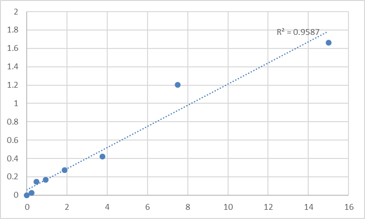
Data standardization and interpretation turn raw absorbance values into biologically meaningful EGF concentrations. First, construct a calibration curve using the kit’s 7 pre-calibrated human EGF standards (0.1–20 ng/mL) and fit with a four-parameter logistic (4PL) regression (R² ≥ 0.995 is mandatory for academic publications)—linear regression underestimates low and high EGF concentrations. Calculate sample EGF levels using the 4PL equation, then normalize to total protein concentration (via BCA assay) for tissue homogenates or cell lysates—express results as “ng/mg protein” for comparative analysis (e.g., tumor vs. normal tissue). For clinical samples, express results as “ng/mL” and compare to established reference ranges (e.g., elevated EGF >5 ng/mL in colorectal cancer patients). Avoid a common pitfall: Never extrapolate beyond the standard curve—dilute high-EGF samples (e.g., EGF-treated cell supernatants) to fit within 0.1–20 ng/mL, as values outside this range are statistically unreliable.
The versatility of EliKine™ Human EGF ELISA Kit KTE6005 aligns with the diverse needs of human EGF research and clinical applications. In oncology, it quantifies EGF in patient serum or tumor lysates to correlate expression with disease stage, prognosis, or response to anti-EGF therapies (e.g., cetuximab). In dermatology, it measures EGF in skin biopsies or wound exudates to evaluate healing efficacy of topical treatments. In regenerative medicine, it monitors EGF secretion in stem cell cultures to optimize tissue engineering protocols. For drug development, it serves as a high-throughput tool to screen compounds that modulate EGF secretion or receptor binding, accelerating preclinical testing of EGF-targeted therapeutics. What sets KTE6005 apart is its cost-effectiveness ($3.94 per test) without compromising performance—making it accessible to academic labs, small biotechs, and clinical diagnostic facilities with limited budgets.
Best practices for kit storage and quality control ensure consistent performance across experiments. Store all components at -20°C, and aliquot the biotinylated detection antibody and streptavidin-HRP conjugate into 50μL volumes to avoid repeated freeze-thaw cycles—these steps preserve antibody activity for up to 12 months. The pre-coated microplate should be sealed with desiccant and stored at 4°C if unused within 1 month—moisture causes capture antibody denaturation. Include a positive control (pooled serum from healthy donors with known EGF levels) and a negative control (EGF-depleted serum) in every assay run to monitor batch-to-batch variability—coefficient of variation (CV) < 8% is acceptable for human EGF quantification. For longitudinal clinical studies (e.g., tracking EGF over 6 months in cancer patients), use the same kit batch to minimize inter-assay variability, a critical factor for detecting subtle, clinically relevant changes.
In conclusion, Abbkine’s EliKine™ Human EGF ELISA Kit KTE6005 delivers the specificity, sensitivity, and cost-effectiveness required for rigorous human EGF quantification. By following tailored sample preparation, optimized assay conditions, interference mitigation, and robust data standardization, researchers and clinicians can generate publication-quality results that advance disease research and therapeutic development. This kit’s professional-grade design, combined with its promoted price point, makes it an indispensable tool for anyone working with human EGF in clinical, academic, or industrial settings. To integrate KTE6005 into your workflow, visit its product page for detailed technical notes and application examples.
Would you like me to create a customized protocol template for your specific sample type (e.g., human serum, tumor lysates, cell culture supernatants) or research application (e.g., cancer biomarker validation, drug screening, wound healing studies) to further optimize EGF quantification with KTE6005?


