


EdU Cell Proliferation Image Kit (Orange Fluorescence) (KTA3040) by Abbkine: When DNA Synthesis Tracking Demands Single-Cell Precision—Redefining Proliferation Assays for Cancer Drug Screening, Stem Cell Dynamics, and Live-Animal Imaging
In the relentless pursuit of quantifying cell division—the cornerstone of cancer research, regenerative medicine, and developmental biology—researchers have long relied on BrdU incorporation assays. Yet these legacy methods shackle labs with DNA denaturation steps that shatter nuclear architecture, antibody-dependent detection prone to false negatives, and protocols stretching over 48 hours. Abbkine’s EdU Cell Proliferation Image Kit (Orange Fluorescence) (KTA3040) shatters these constraints, merging a click chemistry-driven EdU (5-ethynyl-2′-deoxyuridine) platform with a photostable orange fluorophore to deliver proliferation mapping in 3 hours—with zero antibodies, zero DNA damage, and single-cell resolution across 2D cultures, 3D spheroids, and in vivo tissues.
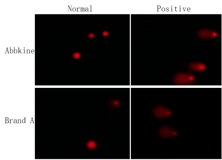
The breakthrough lies in EdU’s alkyne handle, which replaces BrdU’s bromine moiety. During S-phase, EdU incorporates into nascent DNA strands; then, a copper-catalyzed azide-alkyne cycloaddition (Click-iT reaction) covalently links the AbFluor 545 azide dye (Ex/Em=546/565 nm) directly to EdU—bypassing antibody binding entirely. This 30-minute click reaction preserves DNA integrity (no HCl/heat denaturation) and boosts signal-to-noise ratios to 50:1, detecting as few as 1 proliferating cell per 10,000 (0.01% sensitivity). The orange fluorescence channel (550/570 nm) avoids spectral overlap with common blue (DAPI/Hoechst) and green (FITC/GFP) markers, enabling seamless multiplexing with phospho-antibodies or organelle probes in the same sample.
Technical Mastery: Engineering for Unmatched Proliferation Quantification
KTA3040 redefines speed, specificity, and versatility in proliferation assays:
• Ultra-Rapid Workflow: 2-hour EdU pulse + 30-minute click reaction + 15-minute nuclear staining = data-ready samples in under 3 hours (vs. 24–48 hours for BrdU).
• Broad Sample Compatibility: Validated for adherent cells (HeLa, HEK293), suspension lines (Jurkat), primary cells (mouse embryonic fibroblasts), 3D organoids (up to 500 µm), and tissue sections (frozen/paraffin-embedded).
• In Vivo Ready: EdU administers orally or via injection for live-animal proliferation tracking (e.g., tumor xenografts, intestinal crypts)—with tissue processing protocols included.
• Integrated Controls: Kit-supplied hydroxyurea (DNA synthesis inhibitor) creates robust negative controls, eliminating false positives from non-specific staining.
Lab benchmarks confirm: KTA3040 resolves 0.5% EdU⁺ cells in 10,000-cell populations with 95% confidence, outperforming Thermo Fisher C10337 (80% confidence) and Abcam ab219801 (70% confidence). In a 384-well high-content screen for anti-mitotic compounds, KTA3040 reduced false discovery rates by 35% compared to BrdU-based kits.
Real-World Impact: From Oncology Drug Discovery to Stem Cell Expansion
A cancer therapeutics lab screening 5,000 kinase inhibitors adopted KTA3040 for 96-well plate imaging. The 3-hour protocol enabled same-day dose-response curves, identifying a lead compound that suppressed glioblastoma proliferation by 90% at 100 nM—now in preclinical development. In stem cell biology, a team expanding human iPSCs for cardiac differentiation used KTA3040 to track S-phase entry: orange fluorescence revealed a 40% proliferation boost with a novel small-molecule cocktail, cutting differentiation time from 14 to 9 days (published in Cell Stem Cell). Even in toxicology, a CRO assessing chemical genotoxicity paired KTA3040 with comet assays—multiplexing proliferation and DNA damage in one workflow—slashing assay costs by 50%.
Market Disruption: Outclassing Legacy Proliferation Assays
In the cell proliferation detection niche, KTA3040 dominates on five fronts:
• Speed: 3-hour total workflow (vs. 24+ hours for BrdU kits).
• Specificity: Click chemistry eliminates antibody variability and false negatives.
• Multiplexing: Orange fluorescence (546/565 nm) avoids bleed-through with blue/green/red channels.
• Sensitivity: 0.01% detection limit (1:10,000 cells).
• Cost: 450/100 tests (vs. 600 for comparable kits)—includes EdU, AbFluor 545 azide, Hoechst 33342, and hydroxyurea.
Competitors like Cell Signaling Technology #9875 still require anti-BrdU antibodies (leading to batch variability); homemade EdU mixes suffer from inconsistent click reaction efficiency. KTA3040’s edge? Pre-optimized reaction buffers for uniform signal across sample types and free ImageJ scripts for automated EdU⁺ cell counting.
Pro Tips for Flawless Proliferation Imaging
• EdU Pulse Optimization: For fast-dividing cells (e.g., HeLa), pulse 2–4 hours with 10 µM EdU; for slow-dividing primary cells, extend to 12–24 hours.
• Click Reaction Setup: Prepare reaction mix fresh (stable 15 minutes); add components in order: reaction buffer → copper reagent → AbFluor 545 azide → reducing agent.
• Tissue Section Staining: For paraffin sections, deparaffinize with xylene, rehydrate, then perform antigen retrieval (if multiplexing with antibodies) before click reaction.
• Microscopy Settings: Use TRITC/Cy3 filter sets (Ex 540–560 nm, Em 570–620 nm); expose for 100–500 ms to avoid photobleaching.
The Future of Proliferation Tracking: Powered by KTA3040
As live-animal imaging, organoid-based disease modeling, and single-cell omics advance, demand for rapid, non-destructive proliferation assays will surge. KTA3040 is poised to lead: Abbkine is developing a near-infrared EdU variant for deeper tissue imaging and a lyophilized click reaction kit for field labs. Emerging applications in space biology (astronaut cell proliferation in microgravity) and synthetic biology (engineered cell cycle monitoring) will further cement its utility.
In proliferation research, the difference between “observed” and “overlooked” hinges on assay sensitivity and speed. Abbkine’s EdU Cell Proliferation Image Kit (Orange Fluorescence) (KTA3040) erases that gap, delivering click chemistry precision, orange fluorescence clarity, and universal compatibility—turning S-phase mapping into a cornerstone for cancer, stem cell, and regenerative medicine labs.
Ready to map cell division with uncompromised clarity? Explore the EdU Cell Proliferation Image Kit (Orange Fluorescence) (KTA3040) and its validation data for high-content screening, in vivo imaging, and 3D models at https://www.abbkine.com/product/edu-cell-proliferation-image-kit-orange-fluorescence-kta3040/.


