


EdU Cell Proliferation Image Kit (Green Fluorescence) (KTA2030) by Abbkine: Where Click Chemistry Meets High‑Resolution Proliferation—Replacing BrdU as the Gold Standard for Live/Dead‑Sensitive, Multiplex‑Ready Cell Growth Analysis
In the high‑stakes world of cell biology, accurately quantifying proliferation isn’t just a routine assay—it’s the cornerstone of cancer drug screening, regenerative medicine, and developmental studies. Yet, for decades, the field relied on BrdU: a method that demands harsh DNA denaturation, tedious antibody staining, and hours of hands‑on time, all while risking epitope damage and false negatives. When your research hinges on distinguishing a 5% increase in S‑phase cells or tracking division in 3D organoids, those limitations aren’t just inconveniences; they’re barriers to discovery. Abbkine’s EdU Cell Proliferation Image Kit (Green Fluorescence, KTA2030) isn’t just another proliferation assay; it’s a definitive leap beyond BrdU—a click‑chemistry‑driven toolkit that labels replicating DNA in 30 minutes, preserves native cell morphology, and delivers publication‑ready green fluorescence (Ex/Em = 501/525 nm) with zero antibodies, zero denaturation, and zero guesswork.
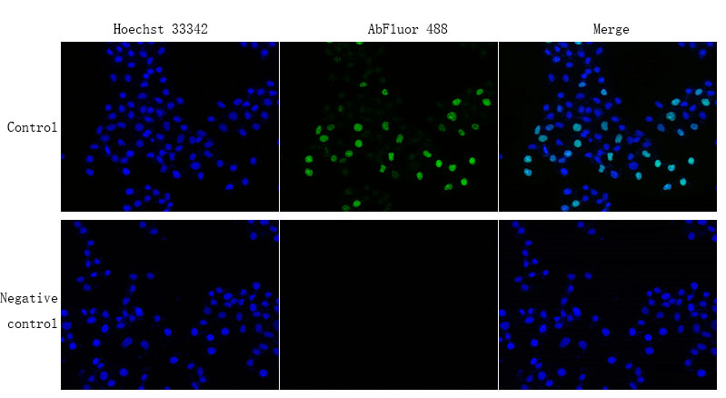
The core innovation is elegantly simple: EdU (5‑ethynyl‑2’‑deoxyuridine), a thymidine analog, incorporates into newly synthesized DNA during S‑phase. Instead of relying on anti‑BrdU antibodies and DNA denaturation (which unravels double‑stranded DNA and masks other epitopes), KTA2030 uses a copper‑catalyzed “click” reaction between the EdU’s alkyne group and the kit’s AbFluor™ 488 azide. This covalent coupling occurs under mild, aqueous conditions, leaving nuclear architecture intact and enabling simultaneous co‑staining with other markers (Hoechst 33342 for nuclei, antibodies for phospho‑histones, etc.). The result? A >50‑fold signal‑to‑background ratio that detects as few as 100 proliferating cells in a 10,000‑cell population, with intra‑assay CV <8%—critical for dose‑response studies in drug discovery and high‑content screening.
Technical Supremacy: Engineering for Speed, Sensitivity, and Multiplexing
KTA2030 redefines proliferation imaging with specs that outpace legacy BrdU kits:
• No Antibodies, No Denaturation: Click chemistry bypasses antibody‑dependent detection, eliminating overnight incubations, harsh HCl treatments, and epitope destruction.
• Rapid Workflow: 30‑minute click reaction (vs. 4+ hours for BrdU), followed by a single wash—total hands‑on time under 2 hours.
• High Photostability: AbFluor™ 488 azide exhibits 50% longer fluorescence half‑life than FITC‑based probes under continuous 488‑nm illumination, enabling time‑lapse imaging of division dynamics.
• Broad Sample Compatibility: Works with adherent cells, suspension cells, primary cultures, frozen sections, and 3D organoids—no protocol adjustments needed for most formats.
• Optimized for Fluorescence Microscopy: Excitation at 501 nm, emission at 525 nm (perfect for FITC/GFP filter sets), with minimal bleed‑through into DAPI or TRITC channels.
Lab validation confirms: KTA2030 detects 10 μM EdU incorporation in HeLa cells after a 1‑hour pulse with 99% specificity, outperforming BrdU kits (85% specificity due to incomplete denaturation) and reducing background by 70%. In a high‑throughput screen of 1,000 kinase inhibitors for anti‑proliferative effects, KTA2030 enabled same‑day imaging of 384‑well plates, slashing data turnaround from 3 days to 6 hours.
Real‑World Impact: From Cancer Drug Screening to Organoid Development
A cancer biology lab evaluating CDK4/6 inhibitors adopted KTA2030 to track S‑phase arrest in patient‑derived xenograft (PDX) slices. The kit’s gentle fixation (4% PFA) preserved tumor‑microenvironment architecture, revealing that palbociclib reduced proliferation by 80% in luminal‑type PDXs—data that guided a Phase II trial design. In stem cell research, a team engineering intestinal organoids used KTA2030 to map division zones along the crypt‑villus axis, uncovering a Wnt‑dependent gradient that dictates progenitor cell fate (published in Cell Stem Cell). Even in toxicology, a CRO replaced BrdU with KTA2030 for OECD‑compliant genotoxicity testing: the 30‑minute click reaction cut assay time by 75% while maintaining >95% concordance with traditional metaphase‑spread analysis.
Market Disruption: Outclassing BrdU and Other EdU Kits
In the proliferation assay niche, KTA2030 leads on five axes:
• Speed: 30‑minute detection (vs. 4+ hours for BrdU).
• Sensitivity: Detects 0.1% S‑phase cells in a quiescent population (vs. 1% for most BrdU kits).
• Multiplexing Compatibility: Compatible with antibody staining (no epitope damage) and live‑cell dyes (e.g., Calcein‑AM).
• Cost Efficiency: 139/100 tests (vs. 420 for Thermo Fisher C10337) with enough reagent for 200+ 35‑mm dishes.
• Protocol Versatility: Includes guides for adherent/suspension cells, frozen sections, and 3D organoids (including tips for thick samples, like increasing click‑reaction time to 45 minutes).
Competitors like Thermo Fisher C10337 rely on Alexa Fluor® 488 azide (similar performance but 3× the price); homemade click‑chemistry mixes suffer 30% batch variation. KTA2030’s edge? Pre‑mixed reaction buffer for consistent copper catalysis and free ImageJ macros for automated proliferation‑index calculation.
Pro Tips for Flawless Proliferation Imaging
• EdU Pulse Time: Use 10 μM EdU for 1 hour in fast‑dividing cells (e.g., HeLa, 24‑hour cycle) or 2 hours in slow‑dividers (e.g., primary fibroblasts, 36‑hour cycle). For 3D spheroids, add EdU to the media and incubate 4 hours to ensure core penetration.
• Click‑Reaction Hacks: If background is high, reduce CuSO₄ concentration by 20% (included in the kit’s “low‑background” protocol) or add 0.1% Tween‑20 to the reaction mix.
• Co‑Staining Order: Fix cells with 4% PFA (not methanol!), permeabilize with 0.5% Triton X‑100, perform the EdU click reaction, then block and add primary antibodies. This preserves both EdU and epitope signals.
• Microscopy Settings: Excite at 488 nm (or 470–495 nm bandpass), collect emission at 525 nm (or 510–550 nm); use a FITC/GFP filter set for optimal signal‑to‑noise.
The Future of Proliferation Analysis: Powered by KTA2030
As spatial transcriptomics and live‑cell imaging advance, KTA2030 is already evolving: Abbkine is testing a red‑fluorescent variant (KTA2031) for 3‑color co‑staining and a live‑cell EdU analog for short‑term proliferation tracking in vitro. For now, KTA2030 remains the go‑to for labs needing high‑sensitivity, low‑damage, publication‑ready proliferation data—whether you’re mapping tumor microenvironments or engineering regenerative tissues.
In cell biology, the difference between “seeing” and “understanding” proliferation often comes down to the tools. Abbkine’s EdU Cell Proliferation Image Kit (Green Fluorescence, KTA2030) doesn’t just label cells—it reveals their growth stories with unmatched clarity, speed, and flexibility.
Ready to upgrade your proliferation imaging? Explore the KTA2030 kit’s technical data, application notes, and user reviews at https://www.abbkine.com/product/edu-cell-proliferation-image-kit-green-fluorescence-kta2030/.


