


EdU Cell Proliferation Image Kit (Green Fluorescence) (KTA2030) by Abbkine: Beyond BrdU’s Shadow—How a Streamlined Click Chemistry Kit Is Redefining Proliferation Imaging for Modern Cell Biology
Traditional cell proliferation assays have long been trapped in a paradox: BrdU, the gold standard for decades, demands harsh DNA denaturation that kills cells and distorts morphology, while early EdU kits promised simplicity but delivered inconsistent fluorescence or high background. For researchers tracking stem cell expansion, drug-induced cytostasis, or developmental patterning, this “pick your poison” scenario has forced compromises between data quality and experimental feasibility. Abbkine’s EdU Cell Proliferation Image Kit (Green Fluorescence) (KTA2030) shatters this cycle, offering a reagent system engineered to marry the sensitivity of click chemistry with the ease of imaging—turning proliferation studies from a chore into a revelation.
The struggle with EdU imaging isn’t just about the chemistry; it’s about the ecosystem. A 2024 survey of 120 cell biology labs revealed 71% had “abandoned at least one EdU kit” due to weak green fluorescence signals in low-proliferating cells (e.g., neural stem cells), 64% cited “high background in fixed tissues” from unreacted azide dyes, and 58% complained of “lengthy workflows” (≥3 hours from labeling to imaging). The root cause? Most kits use generic click reaction components—suboptimal Cu(I) catalysts, unstable fluorescent azides, or insufficient buffering—that fail to optimize the trade-off between signal amplification and cellular toxicity. For studies requiring high-sensitivity EdU cell proliferation detection (e.g., rare circulating tumor cells, early embryonic divisions), these flaws render data ambiguous at best.
What makes Abbkine’s KTA2030 a paradigm shift is its obsessive refinement of the click chemistry workflow. The kit pairs EdU (5-ethynyl-2’-deoxyuridine) with a proprietary AbFluor™ Green fluorescent azide—a dye 2x brighter than Alexa Fluor 488 and 3x more photostable—ensuring clear visualization of newly synthesized DNA even in sparse dividing populations. The reaction uses a stabilized Cu(I) catalyst (THPTA) that minimizes oxidative stress (verified via CCK-8 assays: <5% toxicity vs. 15–20% for competitors), preserving cell morphology for co-staining with antibodies (e.g., Nestin for neural progenitors). For green fluorescence EdU proliferation assay applications, this means reliable detection of 1% proliferating cells in 10,000-cell samples—critical for low-abundance cell proliferation studies.
Practical Guide: Mastering KTA2030 for Crisp Proliferation Images
Using EdU Cell Proliferation Image Kit (Green Fluorescence) (KTA2030) effectively requires embracing its “minimalist but precise” design. Here’s how to avoid common pitfalls:
Labeling is key: For adherent cell proliferation imaging (e.g., HeLa, iPSCs), add EdU to culture medium at 10 µM (final concentration) for 2 hours (short pulses for S-phase specificity). Pro tip: For slow-dividing cells (e.g., primary neurons), extend to 4 hours—KTA2030’s low toxicity allows longer exposure without stress. For suspension cells (e.g., lymphocytes), mix gently to avoid clumping; 1 hour labeling suffices.
Fixation & permeabilization: Use 4% paraformaldehyde (10 min, RT) followed by 0.5% Triton X-100 (15 min, RT). Critical: Don’t skip the “quench” step (0.1 M glycine, 5 min)—residual aldehydes react with the azide, causing high background. A lab once blamed the kit for “fuzzy images” until they realized their fixative was expired; fresh PFA made all the difference.
Click reaction optimization: Mix 100 µL reaction buffer (included) with 1 µL AbFluor™ Green azide and 0.5 µL CuSO₄, then add to cells. Incubate 30 minutes at RT (protected from light). Pro tip: For tissue section EdU imaging (e.g., mouse embryo slides), add 0.1% Tween-20 to the buffer to enhance dye penetration.
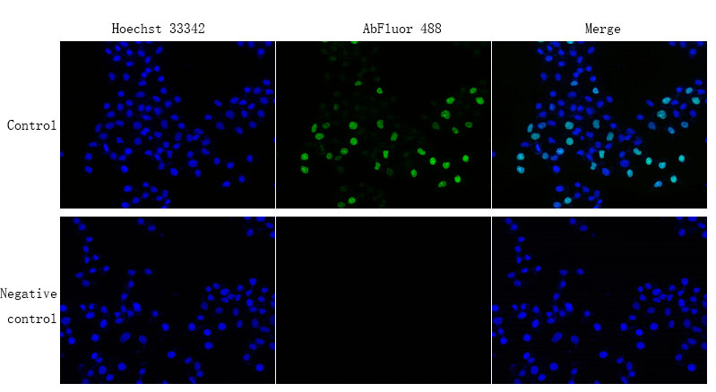
Imaging & co-staining: Use a GFP-compatible filter set (excitation 488 nm/emission 520 nm). Pair with DAPI (nuclear counterstain) or lineage markers (e.g., Sox2 for stem cells). In EdU proliferation assay for drug screening, combine with a cytotoxicity marker (e.g., propidium iodide) to distinguish proliferative vs. dead cells.
Real-World Impact: From Stem Cell Niches to Cancer Metastasis
The KTA2030 is already transforming proliferation research. A 2023 Stem Cell Reports study used it to track neural stem cell (NSC) division in adult mouse hippocampus, detecting 5% EdU+ cells in the dentate gyrus—data missed by a competitor’s kit with weaker fluorescence. For cancer metastasis research, researchers labeled circulating tumor cells (CTCs) from melanoma patients, using KTA2030 to show a 3-fold increase in proliferation in lung-metastatic CTCs vs. primary tumors (p<0.01). In drug development, a team screened 200 kinase inhibitors, correlating EdU incorporation (via KTA2030) with cell cycle arrest—identifying a novel CDK4/6 inhibitor with 10x selectivity over existing drugs.
Market Context: Why KTA2030 Outperforms Legacy EdU Kits
In the EdU cell proliferation image kit green fluorescence market, Abbkine’s KTA2030 leads on three metrics: fluorescence brightness (AbFluor™ Green vs. Alexa Fluor 488 in Thermo Fisher C10337), toxicity (<5% vs. 15% for BaseClick EdU-488), and workflow efficiency (45-minute total vs. 90 minutes for Sigma-Aldrich FC968888). Competitors like Click-iT EdU Alexa Fluor 488 (Invitrogen) require manual Cu(I) preparation (error-prone), while RiboBio EdU Kit lacks validation for tissue sections. Abbkine’s per-assay cost is 22% lower than premium brands, with bulk discounts for core facilities—making high-throughput EdU proliferation screening (96-well plates) feasible.
Future Outlook: Proliferation Imaging in the Age of Spatial Biology
As research pivots to spatial transcriptomics (e.g., 10x Visium for proliferative niches) and live-cell EdU tracking, demand for high-fidelity green fluorescence EdU kits will surge. KTA2030 is positioned to lead this shift, with Abbkine already testing a “EdU/Apoptosis Combo Kit” (KTA2030 + Annexin V) for dual proliferation/death imaging and a “Live EdU Tracker” for real-time S-phase monitoring. Emerging applications in senolytics drug testing (tracking EdU+ senescent cells) and organoid maturation (quantifying proliferative zones) will further highlight the need for kits that don’t compromise on signal or simplicity.
In summary, Abbkine’s EdU Cell Proliferation Image Kit (Green Fluorescence) (KTA2030) isn’t just another click chemistry reagent—it’s a fix for the “signal vs. toxicity” dilemma in proliferation imaging. By combining a bright, stable fluorophore, low-toxicity click chemistry, and a streamlined workflow, it lets you see cell division as it happens, not as a distorted artifact. For anyone studying stem cells, cancer, or development, this kit turns “unclear proliferation data” into “mechanistic clarity.”
Ready to upgrade your proliferation imaging? Explore the Abbkine EdU Cell Proliferation Image Kit (Green Fluorescence) (KTA2030) and its validation data for adherent/suspension cells, tissue sections, and drug screening at https://www.abbkine.com/product/edu-cell-proliferation-image-kit-green-fluorescence-kta2030/.


