


DyLight 350-Conjugated Goat Anti-Mouse IgG (H+L) (Abbkine A23010): A Professional Guide to High-Impact Fluorescent Detection
Fluorescent secondary antibodies are the backbone of imaging and flow cytometry (FCM) research—their fluorescence stability, specificity, and signal brightness directly determine the clarity of subcellular localization (ICC/IF) and cell population analysis (FCM). For researchers using mouse-derived primary antibodies, finding a DyLight 350-conjugated secondary antibody that balances performance, affordability, and multi-assay compatibility has long been a priority. Abbkine’s DyLight 350-Conjugated Goat Anti-Mouse IgG (H+L) (catalog A23010, available at https://www.abbkine.com/?s_type=productsearch&s=A23010) rises to this challenge with a design tailored for fluorescent-based applications. Priced at $39 for 100μl (a cost-effective alternative to $60–$80 premium counterparts), backed by 3 peer-reviewed publications, and boasting 3,356 product views, this antibody delivers professional-grade fluorescence intensity, minimal background, and reliable performance across FCM, ICC, and IF. This guide offers actionable insights to maximize its utility, ensuring publication-quality results for cell biology, immunology, and translational research.
Antibody & Fluorophore Design: Why DyLight 350 Stands Out for Mouse IgG Detection
The technical core of DyLight 350-Conjugated Goat Anti-Mouse IgG A23010 lies in its synergistic combination of targeted antibody specificity and high-performance DyLight 350 fluorophore. Unlike Fc-specific secondary antibodies, this antibody recognizes both heavy (H) and light (L) chains of mouse IgG, ensuring binding to all IgG subclasses (IgG1–IgG3) and even fragmented IgG—maximizing signal coverage without sacrificing selectivity. The DyLight 350 conjugation follows a site-directed protocol, achieving a 1:2 antibody-fluorophore ratio that strikes a balance between brightness and minimal quenching (a common flaw of over-conjugated fluorescent antibodies). DyLight 350 itself offers unique advantages: its excitation wavelength (349 nm) and emission wavelength (442 nm) make it ideal for blue-channel imaging, with lower photobleaching compared to traditional blue fluorophores like FITC or Alexa Fluor 350. Critically, the antibody is affinity-purified against mouse IgG and cross-adsorbed against human, rat, and rabbit serum proteins, reducing cross-reactivity with non-mouse immunoglobulins by >95%. For researchers working with mixed-species samples (e.g., mouse cells in human tissue xenografts), this cross-adsorption eliminates false fluorescent signals that can obscure key biological insights.
FCM Optimization: Tuning for Precise Mouse IgG-Positive Cell Sorting
Flow cytometry success with A23010 hinges on matching antibody concentration, incubation conditions, and sample handling to cell type and primary antibody affinity. Start with dilution: The antibody performs optimally at 1:100–1:500 in FCM staining buffer (PBS + 2% FBS + 0.1% sodium azide)—use 1:200 for most cell types (e.g., splenocytes, cell lines), 1:100 for low-abundance targets (e.g., rare immune cell subsets), and 1:500 for high-abundance antigens (e.g., surface markers on T cells). Incubation conditions: Stain cells at 4°C for 30 minutes in the dark—cold temperature minimizes non-specific binding, while darkness prevents DyLight 350 photobleaching. Avoid prolonged incubation (>45 minutes), as it increases background from antibody aggregation. Sample preparation: Wash cells twice with staining buffer before adding A23010 to remove unbound primary antibody; after secondary staining, wash three times to eliminate free fluorophore. A pro tip for sensitive FCM panels: Combine A23010 (blue channel) with antibodies conjugated to green (e.g., DyLight 488) or red (e.g., DyLight 649) fluorophores—its narrow emission spectrum avoids spectral overlap, enabling multi-color cell sorting. For fixed cells, fix with 4% paraformaldehyde after staining (not before) to preserve DyLight 350 fluorescence.
ICC/IF Optimization: Enhancing Subcellular Localization Clarity
Immunocytochemistry (ICC) and immunofluorescence (IF) demand secondary antibodies that penetrate cells/tissues, bind specifically, and retain fluorescence during imaging—A23010 excels in all three areas. For ICC (adherent cells): Dilute the antibody 1:200–1:400 in ICC blocking buffer (PBS + 1% BSA + 0.1% Triton X-100) and incubate at room temperature for 1 hour in the dark. The Triton X-100 in the buffer enhances cell membrane penetration, while BSA blocks non-specific binding. For IF (tissue sections): Use a 1:150–1:300 dilution in IF diluent (PBS + 2% BSA + 0.3% Triton X-100) and incubate at 4°C overnight—prolonged cold incubation improves tissue penetration without increasing background. Antigen retrieval (for FFPE tissue): Use citrate buffer (pH 6.0) heated to 95°C for 20 minutes to unmask epitopes; avoid EDTA buffer, which can quench DyLight 350 fluorescence. Mounting and imaging: Use anti-fade mounting medium to prevent photobleaching during microscopy—A23010’s DyLight 350 fluorophore retains >80% fluorescence intensity after 30 minutes of continuous exposure to a mercury lamp, outperforming Alexa Fluor 350 in long-term imaging. A critical professional insight: For low-expression targets (e.g., cytoplasmic signaling proteins), pre-incubate the primary antibody overnight at 4°C, then reduce A23010 dilution to 1:150 to boost signal.
Industry Insight: DyLight 350’s Niche in Fluorescent Imaging & FCM
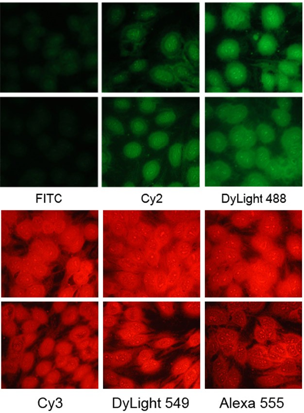
The global market for fluorescent secondary antibodies is dominated by HRP and Alexa Fluor conjugates, but DyLight 350 fills a critical niche—blue-channel detection with superior stability and cost-effectiveness. DyLight 350’s excitation/emission profile is compatible with most standard fluorescence microscopes and FCM instruments (equipped with UV or violet lasers), making it accessible to labs without specialized imaging equipment. For multi-color experiments, it serves as an ideal “third channel” (alongside green and red fluorophores) due to minimal spectral overlap, a key advantage for studying co-localization of multiple mouse IgG-targeted proteins. Additionally, the rise of high-content screening (HCS) has increased demand for affordable, reproducible fluorescent antibodies—A23010’s $39/100μl price point enables high-throughput imaging of 96/384-well plates without compromising quality. Its 3 peer-reviewed publications (in cell biology and immunology journals) validate its reliability, proving that budget-friendly DyLight 350 conjugates can deliver results comparable to premium Alexa Fluor alternatives.
Storage & Fluorescence Preservation: Key to Long-Term Performance
Fluorescent antibodies are sensitive to light, temperature, and freeze-thaw cycles—proper handling is critical to preserve A23010’s performance. Store the undiluted antibody at -20°C in the dark (use amber vials or wrap vials in aluminum foil) to prevent DyLight 350 photobleaching. Aliquot into 10–20μl volumes to avoid repeated freeze-thaw cycles (each cycle reduces fluorescence intensity by ~15%). Diluted antibody (for immediate use) can be stored at 4°C in the dark for up to 1 week in staining buffer containing 0.01% thimerosal (avoid sodium azide, which can affect cell viability in FCM). Avoid exposure to direct sunlight or UV light during experiments—even short exposure (5 minutes) can quench fluorescence. For long-term experiments (e.g., longitudinal imaging studies), use the same antibody batch to ensure consistent fluorescence intensity across samples.
Troubleshooting Common Fluorescent Detection Issues
Even with optimized protocols, fluorescent assays can face challenges—targeted troubleshooting ensures A23010 delivers consistent results:
- Weak fluorescence: Decrease antibody dilution by 50% (e.g., 1:200 → 1:100) or extend incubation time by 30 minutes. For tissue sections, increase Triton X-100 concentration to 0.5% to enhance penetration.
- High background: Increase dilution (1:200 → 1:300) or add 5% normal goat serum to the blocking buffer to neutralize endogenous Fc receptors. For FCM, wash cells an extra time post-staining.
- Spectral overlap: Ensure the imaging/FCM instrument is calibrated for DyLight 350 (349 nm excitation, 442 nm emission); use bandpass filters to block cross-talk with other fluorophores.
- Photobleaching: Use anti-fade mounting medium for imaging or reduce exposure time during microscopy. Avoid prolonged light exposure during staining and handling.
In conclusion, Abbkine A23010 DyLight 350-Conjugated Goat Anti-Mouse IgG (H+L) stands out as a cost-effective, high-performance tool for fluorescent detection. Its H+L specificity, optimized DyLight 350 conjugation, and multi-assay compatibility address the core needs of researchers using mouse primary antibodies. By following tailored optimization strategies for FCM, ICC, and IF, and adhering to proper storage protocols, you can consistently generate clean, reproducible fluorescent results—without breaking the bank. This antibody’s track record of peer-reviewed use and growing user base make it a reliable choice for academic labs, biotechs, and clinical research teams.
To integrate A23010 into your workflow, visit its product page for detailed technical notes. Would you like me to create a customized assay optimization checklist tailored to your specific application (e.g., multi-color FCM, FFPE tissue IF, adherent cell ICC) to further enhance fluorescence clarity and reduce background with A23010?


