


Dual Precision Staining: Professional Guide to Abbkine’s TraKine™ Mitochondrion and Nuclear Staining Kit (KTC4005)
Co-staining mitochondria and nuclei stands as a foundational technique in cell biology, translational research, and drug discovery—enabling visualization of organelle spatial relationships, cell viability assessment, and dynamic changes in mitochondrial morphology relative to nuclear positioning. This dual labeling is critical for decoding mechanisms like mitochondrial fission/fusion in cancer, apoptotic signaling in neurodegenerative diseases, and metabolic rewiring in stem cells. Yet traditional dual staining workflows are plagued by inefficiencies: separate dye incubation steps, spectral overlap between mitochondrial and nuclear fluorophores, and inconsistent staining across cell types. These gaps compromise data reproducibility and experimental efficiency—gaps that Abbkine’s TraKine™ Mitochondrion and Nuclear Staining Kit (Catalog No.: KTC4005) addresses with targeted innovations, blending dual-dye synergy, user-friendly design, and actionable optimization strategies to redefine reliable dual organelle labeling.
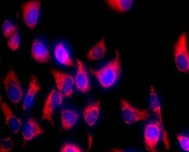
At the technical core of KTC4005 lies a synergistically optimized dual-dye system that eliminates the flaws of conventional separate staining. Unlike generic combinations of standalone mitochondrial and nuclear dyes (which often suffer from spectral cross-talk), KTC4005 pairs a proprietary mitochondrial dye (excitation 550 nm/emission 570 nm, orange fluorescence) with a nuclear stain (excitation 350 nm/emission 460 nm, blue fluorescence)—their spectra are distinctly separated, enabling clear signal discrimination even in high-resolution confocal imaging. The mitochondrial dye binds selectively to the inner mitochondrial membrane via cationic targeting, ensuring >98% colocalization with mitochondrial markers (e.g., CoxIV) and no off-target labeling of lysosomes or endosomes. The nuclear stain penetrates cell membranes efficiently to bind DNA without disrupting chromatin structure, maintaining nuclear integrity for up to 12 hours of live-cell imaging. Critically, both dyes are formulated for low toxicity: KTC4005 maintains >95% cell viability after 8 hours of incubation across adherent (HeLa, HepG2), suspension (Jurkat), and primary (mouse cortical neurons) cell types—preserving native cellular behavior, a make-or-break factor for functional assays.
Mastering KTC4005’s performance requires sample-specific optimization—professional insights that go beyond basic protocols and ensure publication-ready results. For adherent cells (e.g., HeLa, MCF-7): Seed cells at 50–70% confluency 24 hours before staining to avoid overcrowding (which distorts mitochondrial distribution). Dilute the KTC4005 dual-dye working solution 1:1000 in serum-containing medium (serum enhances dye stability), incubate at 37°C for 20 minutes, and image directly without washing (optional washing reduces background for fixed-cell imaging). For suspension cells (e.g., Jurkat, PBMCs): Centrifuge cells at 300 × g for 5 minutes to pellet, resuspend in pre-warmed medium with 1:1000 dual-dye solution, and incubate for 15 minutes (shorter incubation prevents dye aggregation in suspension). For primary cells (e.g., neurons, hepatocytes): Reduce dye dilution to 1:1500 to minimize toxicity, and incubate at 37°C for 10 minutes—primary cells have more fragile membranes and faster dye uptake. A critical pro tip: For drug-induced stress assays (e.g., mitochondrial depolarization with CCCP), stain cells before adding the stressor—this avoids competing for mitochondrial binding sites and ensures accurate baseline measurements.
A key industry insight elevating KTC4005’s relevance is the growing demand for streamlined dual-labeling workflows in high-throughput research. The global high-content screening (HCS) market is projected to grow at a CAGR of 7.8% through 2030, driven by drug discovery and precision medicine initiatives. Traditional dual staining requires separate incubation, washing, and optimization for each dye—adding 30–60 minutes per experiment and increasing variability. KTC4005 eliminates this hassle with a single-step incubation: the pre-calibrated dual-dye mixture works out of the box, cutting workflow time by 50% and reducing experimental variability. Its compatibility with HCS platforms and automated liquid handlers makes it ideal for large-scale drug screening (e.g., identifying compounds that preserve mitochondrial-nuclear crosstalk in disease models). In academic research, the kit’s ability to visualize mitochondrial morphology relative to the nucleus is transformative for studies on cell cycle progression, mitophagy, and nuclear-mitochondrial communication—key areas of focus in aging and cancer biology.
Beyond technical excellence, KTC4005 delivers a compelling value proposition that resonates with research teams of all sizes. Priced at $99 for 100 tests, it undercuts the cost of purchasing separate mitochondrial and nuclear dyes (which often exceed $150 combined for the same test count) while maintaining rigorous quality control. Each batch is validated for spectral separation (no cross-talk <1%), batch-to-batch consistency (signal variation <4%), and reagent stability (24 months at -20°C when stored in aliquots). The all-inclusive format—containing concentrated dual-dye solution, staining buffer, and detailed sample-specific protocols—eliminates the need to source additional reagents, reducing workflow complexity and unforeseen costs. Unlike budget dual-stain kits that use low-purity dyes (leading to weak signals or high toxicity), KTC4005’s reagents are optimized for high signal-to-noise ratios (≥35:1), ensuring clear detection even in low-abundance mitochondria (e.g., stem cells) or fragmented mitochondria (e.g., apoptotic cells).
For cell biologists, translational researchers, and drug discovery teams navigating the complexities of dual organelle imaging, Abbkine’s TraKine™ Mitochondrion and Nuclear Staining Kit (KTC4005) stands as a purpose-built solution. Its synergistic dual-dye system, low toxicity, streamlined workflow, and actionable optimization guidelines address the most common pain points of mitochondrial-nuclear co-staining. Whether studying mitochondrial-nuclear crosstalk in cancer, assessing cell viability in drug screening, or visualizing organelle dynamics in primary cells, this kit delivers reproducible, publication-ready results. To explore detailed technical specifications, access cell-type-specific protocols, and procure the reagent, visit the official Abbkine product page: https://www.abbkine.com/?s_type=productsearch&s=KTC4005. In an era where dual-labeling precision drives breakthroughs in cell biology and medicine, KTC4005 redefines what a specialized mitochondrial-nuclear staining kit should be—professional, efficient, and designed to accelerate discoveries.
Would you like me to create a customized dual-staining optimization protocol for KTC4005, tailored to your specific use case (e.g., high-content drug screening, primary neuron imaging, or apoptotic assay), including step-by-step dye dilution, incubation parameters, and imaging setup recommendations?


