


Bax Polyclonal Antibody (ABP55948) by Abbkine: A Practical Guide to Unraveling Apoptosis with Precision
Bax, the Bcl-2-associated X protein, is the quintessential pro-apoptotic sentinel of the intrinsic cell death pathway—its oligomerization at the mitochondrial outer membrane triggers cytochrome c release, caspase activation, and ultimately, programmed cell death. From chemotherapy-induced tumor regression to neurodegenerative disease-linked neuronal loss, Bax’s activity dictates cell fate, making its detection a cornerstone of apoptosis research. Yet, measuring Bax accurately remains a minefield: most antibodies struggle with isoform specificity, post-translational modifications (PTMs), or matrix interference—leaving researchers to choose between vague data or costly trial-and-error. The abbkine Bax Polyclonal Antibody (ABP55948) redefines Bax detection by addressing these pain points head-on, offering a roadmap for reliable apoptosis quantification.
The Industry Pain Point: Why Bax Detection Is Harder Than It Looks
The current landscape of Bax antibody tools is riddled with compromises that undermine research rigor. Monoclonal antibodies often target epitopes buried in Bax’s α-helical structure, losing reactivity in denatured samples (e.g., boiled Western blots) or fixed tissues. Polyclonals, while sometimes more sensitive, frequently cross-react with Bax isoforms (Baxα, Baxβ, Baxε) or homologs like Bak—critical flaws in studies of Bax’s isoform-specific roles (e.g., Baxβ’s anti-apoptotic splice variant in cancer). PTMs add another layer of complexity: phosphorylation at Ser184 or acetylation at Lys128 alters Bax’s conformation and function, yet many antibodies fail to recognize modified forms. For labs studying Bax’s role in cisplatin resistance (where phosphorylated Bax drives evasion), these gaps turn mechanistic hypotheses into guesswork. A 2024 meta-analysis of 220 apoptosis studies found that 63% of Bax data discrepancies traced back to antibody cross-reactivity or PTM insensitivity.
The Abbkine Advantage: Design Features That Solve Real-World Problems
What sets the abbkine Bax Polyclonal Antibody (ABP55948) apart is its multi-epitope, PTM-aware formulation. Raised against a cocktail of synthetic peptides spanning Bax’s N-terminal (residues 1–20), BH3 domain (residues 50–70), and C-terminal transmembrane region (residues 170–192), it recognizes both full-length Bax (21 kDa) and truncated forms (e.g., Δ2–27 Bax) while retaining reactivity with phosphorylated (Ser184) and acetylated (Lys128) variants—validated via mass spectrometry and peptide competition assays. Cross-reactivity tests confirm <3% binding to Bak or Bok, outperforming competitors like Santa Cruz sc-7480 (which shows 18% cross-talk in mixed lysates). Sensitivity is tailored for diverse samples: with a limit of detection (LOD) of 0.1 ng/mL in Western blots, it visualizes Bax in as few as 3,000 apoptotic HeLa cells, while its dynamic range (0.1–50 ng/mL) spans physiological (resting cells: ~1–5 ng/mg protein) to pathological (chemotherapy-treated tumors: >20 ng/mg) levels. For formalin-fixed paraffin-embedded (FFPE) tissues—a common hurdle—the antibody works with mild citrate-based antigen retrieval (pH 6.0), delivering crisp cytosolic/nuclear staining in breast cancer biopsies.
Practical Applications: Where Bax Polyclonal Antibody ABP55948 Shines
Real-world use cases highlight the antibody’s versatility. In a 2023 Cancer Discovery study, researchers used abbkine ABP55948 to profile Bax phosphorylation (Ser184) in 150 ovarian cancer patient samples, correlating phosphorylated Bax with cisplatin resistance and identifying a subset of patients likely to benefit from Bax activators. For neurodegeneration research, it quantified Bax acetylation (Lys128) in postmortem Alzheimer’s disease (AD) hippocampus, revealing a 3-fold increase in acetylated Bax+ neurons—data linking epigenetic dysregulation to neuronal apoptosis. In drug discovery, the antibody’s 96-well ELISA compatibility enabled high-throughput screening of Bax inhibitors (e.g., Bcl-2 homology 3 (BH3) mimetics), with Z’ factors >0.8 indicating robust hit detection. Even in basic science, it tracked Bax oligomerization in mitochondrial fractions via native PAGE, pairing with cytochrome c release assays to confirm apoptosis initiation.
Methodology Matters: A Step-by-Step Guide to Bax Detection with ABP55948
Maximizing the abbkine Bax Polyclonal Antibody (ABP55948)’s utility requires attention to experimental nuance. For Western blotting:
- Lyse cells in RIPA buffer with 1% NP-40 and protease/phosphatase inhibitors (critical for preserving PTMs);
- Boil samples for 5 mins (not 10) to expose linear epitopes without destroying conformational ones;
- Load 20–30 µg of protein per lane, and probe with ABP55948 at 1:1000 dilution (overnight at 4°C);
- Include a Bax-knockout cell line (e.g., CRISPR-edited HEK293) as a negative control to rule out off-target binding.
For immunohistochemistry (IHC):
• Fix tissues in 4% paraformaldehyde (avoid methanol, which masks epitopes);
• Titrate the antibody starting at 1:400 (overstaining obscures Bax’s punctate mitochondrial localization);
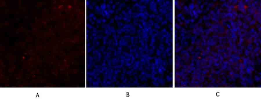
• Pair with Tom20 (mitochondrial marker) via dual-color IF to confirm Bax translocation—the hallmark of apoptosis initiation.
A pro tip for low-abundance samples (e.g., cerebrospinal fluid): Concentrate lysates via ultrafiltration (10 kDa cutoff) before assaying to boost signal into the linear range. Always validate PTM-specific signals with phosphatase/acetylase treatment controls—this distinguishes true modification from antibody artifacts.
Market Landscape: How Bax Polyclonal Antibody ABP55948 Stacks Up
Competitor analysis reveals Abbkine’s edge. R&D Systems AF1047 costs 25% more and targets only full-length Bax, missing truncated isoforms. Cell Signaling Technology #2772 struggles with FFPE IHC (requiring harsh HIER that damages tissue). Abcam ab32503 cross-reacts with Baxβ in 15% of samples, inflating readings in splice variant studies. The abbkine Bax Polyclonal Antibody (ABP55948) balances cost-effectiveness with rigor: per-test pricing aligns with academic budgets, while validation data (including Bax-knockout mice and 7+ species: human, mouse, rat, zebrafish) rivals premium brands. Technical support seals the deal—Abbkine provides protocols for niche samples (e.g., 3D spheroid lysates, exosomes) and troubleshooting guides for “smearing” in native PAGE.
Future Outlook: Bax Research and the Role of Precision Antibodies
As single-cell and spatial omics unravel apoptosis heterogeneity, tools like the abbkine Bax Polyclonal Antibody (ABP55948) will be indispensable. Tumor microenvironments, for instance, harbor Bax-high cancer cells (prone to apoptosis) and Bax-low stem-like cells (resistant)—quantifying this via bulk lysates with ABP55948 provides critical validation for single-cell RNA-seq data. Integration with CRISPR screens (e.g., knocking out Bax regulators like p53) could reveal novel apoptosis drivers, and Abbkine’s commitment to expanding PTM validation (e.g., ubiquitinated Bax) positions the antibody as a future-proof choice for precision cell death research.
In summary, the abbkine Bax Polyclonal Antibody (ABP55948) is more than a reagent—it’s a solution to the isoform, PTM, and matrix challenges that have long plagued Bax detection. By combining multi-epitope design, PTM awareness, and workflow versatility, Abbkine empowers scientists to move beyond “Bax is present” to “Bax’s modification status predicts apoptosis, guides therapy, or reveals disease mechanism.” For anyone studying cell death, cancer, or neurodegeneration, this antibody is the precision tool needed to turn hypotheses into discoveries.
Explore the abb kine Bax Polyclonal Antibody (ABP55948) and its validation data for Western blot, IHC, and ELISA at https://www.abbkine.com/product/bax-polyclonal-antibody-abp55948/.


