


Abbkine PDGFRα Mouse Monoclonal Antibody (7A3) (ABM40334): A Versatile Tool for PDGFRα Research Across Translational and Clinical Science
Platelet-derived growth factor receptor alpha (PDGFRα) is a cell surface tyrosine kinase receptor that’s a linchpin for mesenchymal cell proliferation, organ development, and wound healing—yet its dysregulation and mutation are also driving forces behind a host of human cancers, including gastrointestinal stromal tumors (GIST), non-small cell lung cancer, and gliomas, as well as rare disorders like idiopathic hypereosinophilic syndrome. For researchers spanning cancer biology, developmental biology, and translational medicine, reliable detection of endogenous PDGFRα is non-negotiable—but the field has long grappled with common frustrations: antibodies that cross-react with PDGFRβ, weak signal in formalin-fixed paraffin-embedded (FFPE) tissue, poor cross-species performance, and high non-specific staining in immunofluorescence (IF). The Abbkine PDGFRα Mouse Monoclonal Antibody (7A3) (Cat. No. ABM40334) (product link: https://www.abbkine.com/product/pdgfr%ce%b1-mouse-monoclonal-antibody-7a3-abm40334/) cuts through these pain points: a mouse-derived, affinity-purified monoclonal antibody engineered against a unique PDGFRα epitope (amino acids 1010–1090), validated for specific detection of human, rat, and mouse PDGFRα across Immunohistochemistry (IHC) and Immunofluorescence (IF)—the two most critical applications for PDGFRα spatial and functional research. This deep dive into ABM40334 breaks down its core design strengths, application-specific optimization, and real-world research value, explaining why it’s quickly become a go-to reagent for labs studying PDGFRα’s role in health and disease.
Targeted epitope selection is the backbone of Abbkine PDGFRα Mouse Monoclonal Antibody (7A3) ABM40334’s unrivaled specificity for endogenous PDGFRα. Unlike generic PDGFRα antibodies that target highly conserved kinase domains shared with PDGFRβ (a common source of cross-reactivity), ABM40334 is raised against a synthetic peptide from the 1010–1090 amino acid region of human PDGFRα—a unique stretch of the protein that eliminates off-target binding to other tyrosine kinase receptors, including PDGFRβ. The antibody is affinity-purified from mouse ascites using this epitope-specific immunogen, a rigorous process that enriches for high-affinity antibody clones and strips out non-specific immunoglobulins—resulting in a reagent that exclusively detects endogenous PDGFRα at its expected molecular weight of 180kD, with no smearing or false bands in complex tissue lysates or sections. This level of specificity is a game-changer for cancer research, where distinguishing PDGFRα from PDGFRβ expression is critical: the two receptors have distinct roles in tumor angiogenesis and progression, and cross-reactive antibodies can skew data on PDGFRα-targeted therapy efficacy. For researchers, this means no more validating band or staining identity—ABM40334 delivers unambiguous PDGFRα detection every time.
Cross-species validation for human, rat, and mouse PDGFRα is a transformative feature of ABM40334 that addresses a core gap in translational PDGFRα research. Most PDGFRα antibodies on the market are validated for just one species, forcing labs to purchase and test separate reagents for in vitro cell models (human), in vivo preclinical studies (mouse/rat), and clinical sample analysis (human)—a costly, time-consuming process that introduces experimental variability. ABM40334 is fully validated for specific PDGFRα detection across all three species, with no major protocol tweaks needed for each. This seamless cross-species performance is invaluable for translational research: for example, a lab studying PDGFRα mutations in GIST can use ABM40334 to profile PDGFRα expression in mouse GIST models, then apply the exact same antibody to human FFPE GIST tissue samples—ensuring direct comparability between preclinical and clinical data. It also eliminates the logistical hassle of managing multiple antibodies, a huge win for small to mid-sized labs with limited reagent budgets.
Immunohistochemistry (IHC) is the cornerstone application for PDGFRα tissue profiling—especially in cancer pathology—and ABM40334 is fine-tuned for consistent, high-performance staining in FFPE sections, the gold standard for archival and clinical tissue samples. The antibody comes with a suggested starting dilution of 1:100–200 for IHC, and real-world validation (in rat spleen FFPE tissue) shows 1:200 is the optimal working dilution for sharp, specific staining with zero non-specific background. The single most important step for IHC success with ABM40334 is antigen retrieval: use sodium citrate buffer (pH 6.0) heated to >98°C for 20 minutes (heat-induced epitope retrieval, HIER)—this mild retrieval method unmaskes the PDGFRα epitope without damaging tissue morphology or denaturing the antibody’s binding site, a balance harsher buffers (e.g., Tris-EDTA pH 9.0) fail to strike. After retrieval, cool sections naturally (don’t rush this step—rapid cooling causes tissue detachment), block with 5% normal goat serum (NGS) + 0.1% Triton X-100 for 1 hour at room temperature, and incubate ABM40334 at 4°C overnight. A 1:200 secondary antibody dilution (room temperature, 30 minutes) is all it takes for strong, specific signal, whether using DAB chromogenic detection or fluorescent IHC. For thick tissue sections (e.g., tumor biopsies, organ sections), drop the dilution to 1:100 to ensure full antibody penetration; for thin sections, 1:200 is perfect for minimizing background.
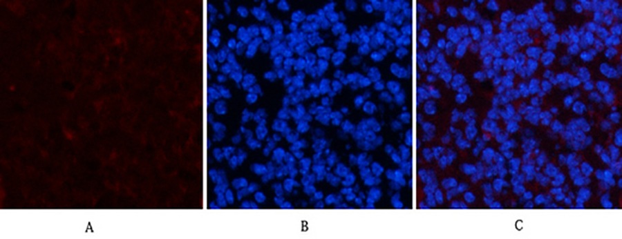
Immunofluorescence (IF) unlocks the spatial localization of PDGFRα in cells and tissues—an essential piece of the puzzle for understanding its functional role—and ABM40334 delivers crisp, non-diffuse staining that makes subcellular and tissue-level PDGFRα mapping straightforward. The antibody’s suggested IF starting dilution is 1:50–200, with 1:200 emerging as the sweet spot for most samples (validated in mouse spleen tissue) for bright, specific red fluorescence (paired with Cy3-conjugated secondary antibodies) and minimal background noise. Unlike some PDGFRα antibodies that produce fuzzy, spread-out staining, ABM40334 labels PDGFRα with sharp, defined signal—critical for co-localization studies with other cell markers (e.g., mesenchymal stem cell markers, tumor angiogenesis markers, or immune cell markers). The IF protocol for ABM40334 is intuitive: fix fresh/frozen sections or cultured cells with 4% paraformaldehyde (PFA) for 15 minutes at room temperature (prolonged fixation masks the PDGFRα epitope), permeabilize with 0.1% Triton X-100 for 10 minutes, block with 5% NGS for 1 hour, and incubate the antibody at 4°C overnight. After washing, use a Cy3-labeled secondary antibody at 1:300 for 50 minutes in the dark, stain nuclei with DAPI, and mount with anti-fade medium—this last step is non-negotiable, as it preserves the fluorescent signal for long-term imaging and analysis. ABM40334’s IF performance also makes it ideal for multi-color staining experiments, with no spectral overlap with common fluorophores (FITC, Alexa Fluor 488, 647), a key feature for modern spatial biology research.
Maximizing the performance and shelf life of ABM40334 boils down to simple, formulation-specific handling and storage practices—something that’s often overlooked but makes a huge difference in experimental reproducibility. The antibody is supplied as a ready-to-use liquid solution at 1 mg/ml, formulated in PBS with 50% glycerol, 0.5% BSA, and 0.02% sodium azide: the glycerol prevents ice crystal formation during freezing (a major cause of antibody denaturation), BSA acts as a protein stabilizer, and sodium azide is a mild preservative safe for all in vitro IHC/IF applications. ABM40334 is stable for one year at -20°C from shipment, but the single most important rule is to aliquot the 30μl stock into 5–10μl volumes upon first use—repeated freezing and thawing is the fastest way to destroy the antibody’s binding affinity. When ready to use, thaw an aliquot on ice (never at room temperature) and centrifuge the vial at 12,000×g for 1 minute before removing the cap—this collects any antibody that’s adhered to the vial wall or lid, ensuring accurate dilutions every time. Prepare fresh antibody dilutions for each experiment (in blocking buffer) and discard any unused diluted antibody—diluted ABM40334 isn’t stable for long-term storage, even at 4°C. For short-term use (1–2 weeks), store thawed aliquots at 4°C; avoid leaving the antibody at room temperature for more than 24 hours, as this leads to rapid degradation.
ABM40334 isn’t just a high-quality PDGFRα antibody—it’s a cost-effective, versatile tool that powers groundbreaking research across multiple high-impact fields, and its value aligns perfectly with the latest industry trends in PDGFRα science. At $109 for 30μl of 1mg/ml antibody, ABM40334 offers unbeatable bang for the buck: the high working dilutions (1:100–200 for IHC/IF) mean a single vial is enough for dozens of experiments, far outperforming more expensive PDGFRα antibodies on the market. Its research applications span the spectrum: cancer biologists use it to profile PDGFRα expression and mutation in GIST and lung cancer samples, developmental biologists study its role in organogenesis and mesenchymal cell differentiation, and translational researchers validate it as a potential diagnostic marker for PDGFRα-driven diseases. What’s more, ABM40334’s focus on IHC and IF— the two most widely used applications for clinical and preclinical tissue research—makes it a practical choice for labs that don’t need Western Blot capability, cutting down on unnecessary reagent costs. As PDGFRα continues to emerge as a key therapeutic target for cancer and rare diseases (with more PDGFRα inhibitors in clinical trials every year), the demand for reliable, specific PDGFRα detection reagents like ABM40334 is only set to grow.
In the end, Abbkine PDGFRα Mouse Monoclonal Antibody (7A3) ABM40334 stands out in a crowded market because it’s designed for real researchers—addressing the actual pain points of PDGFRα research, delivering consistent performance across key applications, and fitting into the budgets and workflows of labs of all sizes. Its targeted epitope design eliminates cross-reactivity, its cross-species validation streamlines translational research, its IHC/IF optimization makes it easy to use with minimal trial and error, and its robust formulation ensures long-term reliability. Whether you’re mapping PDGFRα expression in mouse developmental models, profiling human tumor biopsies for clinical research, or studying PDGFRα’s role in wound healing and tissue regeneration, ABM40334 delivers the specific, reproducible PDGFRα detection you need to generate publication-quality data. For anyone working on PDGFRα—from basic science to translational medicine—this antibody isn’t just a reagent; it’s a research partner that unlocks new insights into one of the most important receptors in health and disease (product link: https://www.abbkine.com/product/pdgfr%ce%b1-mouse-monoclonal-antibody-7a3-abm40334/).


