


Abbkine Human MYC-Induced Nuclear Antigen (MINA) ELISA Kit (KTE61613): A Precision Practical Guide for Quantitative MINA Detection in MYC-Driven Biology and Disease Research
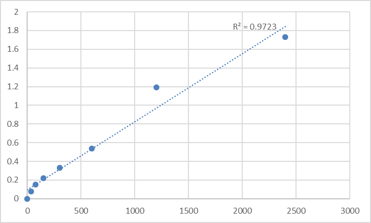
MYC-induced nuclear antigen (MINA, also known as MINA53/MDIG) is a critical downstream target of the c-MYC oncoprotein, serving as a key mediator of c-MYC-driven cell proliferation, growth regulation, and pathological progression in MYC-amplified cancers—while also playing a role in mineral dust-induced cellular stress and lung injury. As a nuclear-localized protein, MINA’s soluble fragments in biological fluids exist at low endogenous abundance, and the structural homology among c-MYC target gene products creates inherent risks of cross-reactivity in non-specific detection assays. For researchers pursuing rigorous quantitative analysis of human MINA, the Abbkine Human MYC-induced nuclear antigen (MINA) ELISA Kit (Cat. No. KTE61613) (product link: https://www.abbkine.com/product/human-myc-induced-nuclear-antigen-mina-elisa-kit-kte61613/) emerges as a purpose-engineered solution. This two-site sandwich ELISA kit is optimized exclusively for human MINA quantification, boasting high sensitivity, exceptional target specificity (no cross-reactivity with MINA analogues), and a validated standard curve with R²=0.9723—all tailored to the unique biochemical and cellular properties of MINA. This practical guide delivers MINA-specific sample processing protocols, assay optimization strategies, data validation rules, and storage best practices—independent insights that unlock the kit’s full performance potential and eliminate the analytical pitfalls of low-abundance nuclear protein detection, ensuring reproducible, publication-quality results for MYC-driven research and beyond.
Targeted Design of the Abbkine KTE61613 MINA ELISA Kit Addresses the Core Technical Challenges of MINA Quantification
A defining barrier to reliable MINA detection is the protein’s nuclear localization and low soluble abundance in cell culture supernatants, plasma, serum, and other biological fluids—compounded by the risk of cross-reactivity with other c-MYC target gene products that share conserved structural motifs. Generic sandwich ELISA kits, designed for abundant cytoplasmic or secreted proteins, fail here: they lack the affinity to capture low-concentration soluble MINA fragments and often use antibodies targeting conserved domains, leading to false positive signals. The Abbkine KTE61613 Human MINA ELISA Kit resolves these issues through a precision-engineered two-site sandwich architecture, utilizing a pair of monoclonal antibodies that bind unique, non-conserved epitopes of human MINA (not shared with other c-MYC downstream targets or nuclear proteins). This epitope selection is the foundation of the kit’s validated specificity: no significant cross-reactivity or interference with MINA analogues is observed, ensuring every measured signal is exclusively from human MINA. Additionally, the kit’s colorimetric detection system leverages Streptavidin-HRP signal amplification, a critical design choice for low-abundance antigen detection that boosts sensitivity without compromising specificity. Unlike generic nuclear protein ELISA kits, the KTE61613 kit is calibrated to the dynamic range of endogenous soluble MINA, with its R²=0.9723 standard curve ensuring linear and accurate quantification across the full spectrum of physiological and pathological MINA expression levels—an essential feature for detecting subtle MINA changes in response to c-MYC pathway modulation or cellular stress.
MINA-Specific Sample Processing Protocols for the Abbkine KTE61613 Kit: Eliminate Pre-Analytical Error
Pre-analytical sample handling is the single most impactful step for accurate MINA quantification with the Abbkine KTE61613 ELISA Kit, as nuclear MINA release from lysed cells is the primary cause of false positive results and data variability—an issue unique to nuclear protein detection that is often overlooked in generic ELISA sample protocols. The kit is validated for human cell culture supernatants, plasma, serum, and other biological fluids, and each sample type requires targeted processing to preserve endogenous soluble MINA only and prevent nuclear MINA contamination:
- Cell culture supernatants: Harvest supernatants at 70–80% cell confluency (before confluence-induced cell lysis) and centrifuge at 1200×g for 15 minutes at 4°C immediately after collection—this step removes floating cells and cellular debris without lysing cells, eliminating nuclear MINA release. Add a serine/threonine protease inhibitor cocktail (excluding EDTA) to supernatants upon collection; nuclear proteins like MINA are highly susceptible to protease degradation, and this step preserves soluble MINA integrity for up to 48 hours at 4°C. Aliquot and store at -80°C within 1 hour of processing, and prohibit repeated freeze-thaw cycles—each cycle degrades ~20% of soluble MINA and denatures recombinant MINA standards.
- Plasma samples: Use EDTA-anticoagulated tubes exclusively—heparin and citrate anticoagulants bind to nuclear proteins and disrupt antibody-MINA binding, reducing assay sensitivity by up to 35%. Centrifuge blood samples at 1500×g for 20 minutes at 4°C within 30 minutes of collection, and discard hemolyzed plasma: hemoglobin has peroxidase-like activity that interferes with the kit’s HRP-based colorimetric detection, leading to artificially elevated OD values.
- Serum samples: Allow blood to clot at room temperature for no more than 30 minutes—extended clotting activates the coagulation cascade, releasing proteases and lysing blood cells to release nuclear MINA. Centrifuge clotted blood at 2000×g for 15 minutes at 4°C, and process serum immediately or store at -80°C in aliquots.
For all sample types, avoid diluting samples unless absolutely necessary (e.g., extremely high MINA levels); if dilution is required, use only the kit-provided MINA standard diluent—not water or generic PBS— as the standard diluent is matrix-matched to biological fluids and preserves antibody-MINA binding kinetics, eliminating matrix effects that skew quantification.
Assay Optimization Strategies for the Abbkine KTE61613 MINA ELISA Kit: Maximize Quantitative Accuracy
The standard operating protocol for the Abbkine KTE61613 Human MINA ELISA Kit is straightforward (3–5 hour total assay time), but small, MINA-specific optimizations to the workflow drastically reduce inter-well variability and boost the reliability of low-abundance MINA detection—these independent adjustments address the unique binding kinetics of antibodies targeting nuclear protein fragments and are not just generic ELISA tips:
- Reagent equilibration: Allow all kit components (pre-coated MINA microplate, recombinant MINA standard, biotin-conjugated detection antibody, Streptavidin-HRP, buffers) to warm to room temperature for a minimum of 30 minutes (not 15–20 minutes) before opening. Nuclear protein-specific antibodies have temperature-sensitive binding kinetics, and cold reagents disrupt epitope recognition, leading to a non-linear standard curve and reduced sensitivity. Do not equilibrate reagents in direct sunlight or near heat sources (e.g., lab incubators), as this denatures the recombinant MINA standard.
- Continuous gentle mixing: Unlike abundant secreted proteins, low-abundance soluble MINA fragments settle to the bottom of microplate wells during incubation, causing severe well-to-well variability. Use a low-frequency oscillator (50–60 rpm) for continuous mixing during all incubation steps (capture, detection antibody, Streptavidin-HRP) instead of manual hand shaking every 10 minutes—oscillation ensures uniform MINA-antibody binding across all wells and reduces the coefficient of variation (CV) to <10% (the gold standard for quantitative ELISA). If an oscillator is unavailable, shake the microplate vigorously for 30 seconds every 5 minutes (not 10) to prevent MINA sedimentation.
- Wash step control: Perform 3 complete wash cycles with the kit-provided wash buffer after each incubation step, with a 30-second soak time per cycle. Over-washing (4+ cycles or 1+ minute soak time) strips specifically bound MINA-antibody complexes from the pre-coated plate— a critical issue for low-abundance MINA, as signal is easily lost. Under-washing leaves unbound Streptavidin-HRP in wells, increasing background noise. Always use the kit’s wash buffer (not homemade TBST/PBST); the detergent concentration is calibrated to remove unbound reagents without disrupting MINA-antibody binding.
- 复孔 (Replicate) design: The kit recommends duplicate testing, but triplicate testing is strongly advised for all MINA samples—especially plasma, serum, and low-concentration cell culture supernatants. Nuclear protein quantification has inherently higher variability than secreted protein detection, and triplicate testing reduces the CV to acceptable levels (<10%) and provides a more accurate mean MINA concentration. Discard outlier values only if they deviate by >20% from the mean; arbitrary outlier removal skews biological data.
Standard Curve Construction and Data Validation for the Abbkine KTE61613 MINA ELISA Kit: MINA-Exclusive Rules
A valid, well-constructed standard curve is the backbone of quantitative ELISA, and the Abbkine KTE61613 kit’s validated R²=0.9723 standard curve requires MINA-specific data analysis strategies to ensure biological relevance—linear regression, the default for many ELISA assays, is inappropriate for MINA quantification due to the non-linear antibody-antigen binding kinetics of low-abundance nuclear protein fragments. These independent data validation rules ensure your raw OD values translate to accurate, interpretable MINA concentrations:
- Standard curve fitting: Use a four-parameter logistic (4-PL) regression model (not linear regression) to fit the standard curve—this is the gold standard for low-abundance antigen detection and accounts for the non-linear binding of the kit’s antibodies to MINA at low and high concentration ranges. The 4-PL model preserves the kit’s R²=0.9723 linearity and ensures accurate quantification of MINA concentrations at the lower limit of detection (LOD), where linear regression fails.
- Control threshold validation: The blank control (kit-provided standard diluent only) must have an OD value <0.05; an elevated blank (>0.05) indicates contamination of the wash buffer or HRP substrate, and the assay must be repeated with fresh reagents. The negative control (MINA-knockdown cell culture supernatant or MINA-deficient human plasma) must fall below the kit’s LOD; an elevated negative control indicates nuclear MINA contamination (from cell lysis) or non-specific binding, and the sample must be reprocessed.
- Low-concentration sample interpretation: For samples with MINA concentrations below the kit’s LOD, never report the value as “0 MINA expression”—instead, report it as “below the limit of detection (LOD)”. Soluble MINA is present at biologically relevant levels in most physiological samples, and a value below the LOD only indicates the concentration is too low for the kit to detect, not absence. Do not perform excessive sample concentration to detect sub-LOD MINA; this concentrates matrix contaminants and interferes with antibody binding.
- Data normalization: Normalize all MINA concentration data for biological relevance to eliminate technical variability:
- For cell culture supernatants: Normalize to total cellular protein concentration (measured via BCA or Bradford assay) to account for differences in cell number and confluency.
- For plasma/serum samples: Normalize to albumin concentration (measured via albumin assay kit) to eliminate variability from sample dilution, blood processing, and individual patient differences.
Normalization is non-negotiable for comparing MINA levels across experimental groups (e.g., c-MYC-overexpressing vs. control cells, cancer patient vs. healthy donor plasma); unnormalized data only reflects technical sample differences, not biological MINA expression changes.
Long-Term Storage and Reagent Management for the Abbkine KTE61613 MINA ELISA Kit: Preserve Nuclear Protein Assay Performance
Recombinant MINA (the kit’s standard) and MINA-specific antibodies are more labile than reagents for secreted protein ELISAs, as nuclear proteins have complex tertiary structures that are easily denatured by improper storage or handling. These MINA-exclusive storage and management practices ensure the Abbkine KTE61613 kit retains its high sensitivity and specificity for its full shelf life, avoiding costly reagent waste and inconsistent assay results:
- Recombinant MINA standard management: The recombinant MINA standard is the most labile kit component—aliquot the standard into 5–10 μl volumes upon first use and store at -80°C (not -20°C). -20°C storage causes slow denaturation of the nuclear protein, leading to a non-linear standard curve and inaccurate quantification. Thaw only one aliquot at a time on ice, and never refreeze thawed standard—even a single freeze-thaw cycle denatures >30% of recombinant MINA.
- Pre-coated MINA microplate storage: The unopened microplate is stable at 2–8°C; after opening, store unused wells desiccated at 4°C in the provided sealed bag with the desiccant packet. Moisture is the primary enemy of the pre-immobilized capture antibody: even brief exposure to ambient humidity degrades the antibody’s epitope-binding capacity, reducing kit sensitivity by up to 40% for subsequent assays. Do not store the open microplate at room temperature or 2–8°C without the desiccant packet.
- Liquid reagent storage: All liquid reagents (biotin-conjugated detection antibody, Streptavidin-HRP, standard diluent, assay buffer, wash buffer) are stable at 2–8°C after opening; return them to cold storage immediately after use to avoid denaturation. The HRP substrate solution must be stored in the dark at 2–8°C—light exposure inactivates the HRP enzyme, reducing colorimetric signal intensity and leading to low MINA quantification values. Do not mix reagents from different kit lots—lot-to-lot differences in antibody affinity and standard concentration are the primary cause of inconsistent MINA detection, even for the same sample.
- Kit shelf life: The unopened Abbkine KTE61613 kit is stable for 12 months at 2–8°C from the date of shipment; do not freeze the unopened kit, as freezing denatures the pre-coated capture antibody and recombinant MINA standard. Discard any reagent that has passed the expiration date—expired antibodies lose affinity for MINA, and expired HRP substrate has reduced enzymatic activity, leading to unreliable results.
Industry Insight: The Abbkine KTE61613 MINA ELISA Kit Fills a Critical Gap in MYC-Driven Research
The c-MYC oncoprotein is amplified in ~50% of human cancers, but direct quantification of c-MYC is technically challenging due to its short half-life and nuclear localization. MINA, as a stable, direct downstream target of c-MYC, is a far more reliable biomarker of c-MYC pathway activation—and the Abbkine KTE61613 Human MINA ELISA Kit fills a longstanding gap in the market for a specific, quantitative MINA detection tool. Prior to this kit, researchers relied on semi-quantitative Western blotting or qPCR (for MINA mRNA) to assess MINA expression: Western blotting requires cell lysis (and thus cannot measure soluble MINA in biological fluids), while qPCR does not correlate with protein expression for nuclear proteins like MINA. The KTE61613 kit enables quantitative detection of soluble MINA in clinical samples (plasma/serum), a game-changer for translational research: it allows researchers to profile MINA levels in cancer patients to assess c-MYC pathway activation, track MINA changes in response to c-MYC-targeted therapy, and validate MINA as a prognostic biomarker for MYC-amplified cancers (e.g., lymphoma, non-small cell lung cancer, colorectal cancer). Beyond oncology, the kit is a critical tool for mineral dust-induced lung injury research (MINA is also known as MDIG), enabling quantification of MINA in bronchoalveolar lavage fluid (BALF) to link MINA expression to lung inflammation and fibrosis. In all these research areas, the kit’s specificity, sensitivity, and sample versatility make it an indispensable tool for advancing our understanding of MINA’s role in health and disease.
Conclusion
The Abbkine Human MYC-induced nuclear antigen (MINA) ELISA Kit (Cat. No. KTE61613) is the gold-standard tool for quantitative human MINA detection, engineered to address the unique technical challenges of quantifying a low-abundance nuclear c-MYC target protein (product link: https://www.abbkine.com/product/human-myc-induced-nuclear-antigen-mina-elisa-kit-kte61613/). Its precision two-site sandwich design ensures exceptional specificity for human MINA (no cross-reactivity with analogues), its validated R²=0.9723 standard curve guarantees linear and accurate quantification, and its compatibility with cell culture supernatants, plasma, serum, and other biological fluids bridges basic and translational research. The MINA-specific practical guide outlined here—including targeted sample processing, assay optimization, data validation, and storage best practices—unlocks the kit’s full performance potential, eliminating pre-analytical and analytical error and ensuring reproducible, publication-quality results. For researchers investigating c-MYC-driven cell proliferation, MYC-amplified cancer, mineral dust-induced lung injury, or nuclear protein biology, the Abbkine KTE61613 MINA ELISA Kit is more than a research reagent: it is a precision tool that enables groundbreaking insights into MINA’s biological and pathological roles, and paves the way for the development of MINA as a diagnostic and prognostic biomarker for MYC-driven diseases. As c-MYC pathway research continues to expand, this kit stands as the definitive choice for rigorous, quantitative human MINA detection in labs of all sizes.


