


Abbkine GFAP Monoclonal Antibody (ABM0021): Practical Guide for Astrocyte Marker Detection
Glia Fibrillary Acidic Protein (GFAP) is the gold-standard marker for mature astrocytes, central to CNS homeostasis, neuroinflammation, and neurodegenerative disease pathogenesis. Reliable endogenous GFAP detection is critical for neurobiology and neuropathology research, yet the field faces pitfalls like poor cross-species reactivity, inconsistent application performance, and vague optimization guidelines. The Abbkine GFAP Monoclonal Antibody (Cat. No. ABM0021) (product link: https://www.abbkine.com/product/gfap-monoclonal-antibody-abm0021/) solves these issues: a mouse-derived, affinity-purified antibody validated for human, rat, and mouse GFAP across Western Blot (WB), Immunohistochemistry (IHC), and Immunofluorescence (IF). This condensed practical guide focuses on application-specific optimization, antibody handling, and troubleshooting to help researchers generate reproducible, publication-quality results.
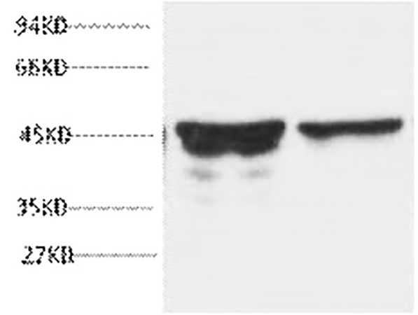
WB Optimization for Abbkine GFAP Monoclonal Antibody ABM0021
The Abbkine GFAP Monoclonal Antibody ABM0021 is validated for WB with a 1:2000–5000 starting dilution, optimized for GFAP’s intermediate filament structure. For CNS tissue (brain/spinal cord), use RIPA buffer with protease inhibitors to preserve GFAP integrity; rat brain tissue performs best at 1:5000, yielding a sharp 45kD band with no off-target signal. For low-GFAP peripheral tissues, use 1:2000 and 20–30μg protein per lane. Key steps: 10% SDS-PAGE gel, PVDF membrane transfer (300mA, 90min, 4°C), 5% milk blocking (1hr, RT), primary incubation (1:2000–5000, 4°C overnight), and anti-mouse IgG HRP-conjugate (1:5000, 1hr, RT). The antibody detects all endogenous GFAP isoforms, critical for studying isoform switching in CNS disease.
IHC Protocol for Abbkine GFAP Monoclonal Antibody ABM0021
IHC success with GFAP Monoclonal Antibody ABM0021 depends on antigen retrieval: use sodium citrate buffer (pH 6.0) at >98°C for 20min (HIER) for paraffin sections—harsh buffers damage GFAP epitopes. Cool sections naturally, block with 5% NGS + 0.1% Triton X-100 (1hr, RT), and incubate primary antibody at 1:50–300 (1:200 optimal) at 4°C overnight. Wash 3x with PBS, incubate secondary antibody (1:200, 30min, RT), and use standard DAB or fluorescent detection. For thick CNS sections (20–30μm), use 1:50; thin sections (5–10μm) use 1:300. Validated for human, mouse, and rat tissues (including peripheral organs), it enables research on peripheral astrocyte-like cells.
IF Optimization for Abbkine GFAP Monoclonal Antibody ABM0021
For IF, GFAP Monoclonal Antibody ABM0021 uses a fixed 1:200 starting dilution. Fix fresh/frozen sections with 4% PFA (15min, RT)—prolonged fixation masks epitopes. Permeabilize with 0.1% Triton X-100 (10min, RT), block with 5% NGS (1hr, RT), and incubate primary antibody at 4°C overnight. Use Cy3-conjugated secondary antibody (1:300, 50min, dark) and DAPI for nuclei; anti-fade mounting medium prevents signal loss. The antibody produces clear filamentous staining, avoiding diffuse background common with low-quality GFAP antibodies, and is compatible with multi-color IF (NeuN, Iba1) for astrocyte crosstalk studies.
Antibody Handling & Storage for ABM0021
Preserve GFAP Monoclonal Antibody ABM0021 (1mg/ml, PBS + 50% glycerol) by aliquoting into 5–10μl volumes upon first use—repeated freeze-thaw denatures the antibody. Store at -20°C (1-year shelf life); thaw on ice and centrifuge (12,000×g, 1min) before use. Prepare fresh dilutions for each experiment; discard unused diluted antibody. Short-term storage (1–2 weeks) at 4°C is acceptable; avoid room temperature storage >24hr. Sodium azide preservative is safe for in vitro use.
Troubleshooting Common Issues
Targeted fixes for GFAP Monoclonal Antibody ABM0021: 1) No signal: Reduce antibody dilution (1:2000 WB) or re-perform HIER (IHC). 2) High background: Shorten blocking (30min WB) or reduce secondary antibody concentration (1:200 IHC/IF). 3) Non-specific CNS staining: Add 0.05% Tween-20 to primary buffer. 4) Distorted filaments: Reduce PFA fixation (10min) or Triton X-100 (0.05%).
Cross-Species Detection Tips
GFAP Monoclonal Antibody ABM0021 reacts with human, rat, and mouse GFAP. Optimal dilutions: Human (1:200 WB/IF, 1:100 IHC), rat (1:5000 WB, 1:200 IHC/IF), mouse (1:3000 WB, 1:200 IHC/IF). No major protocol changes are needed—eliminating the cost of separate species-specific antibodies for translational research.
Conclusion
Abbkine GFAP Monoclonal Antibody (ABM0021) is the gold-standard for GFAP detection across WB, IHC, and IF. Its cross-species reactivity, specificity, and consistent performance make it indispensable for astrocyte biology, neuroinflammation, and neurodegenerative disease research. This practical guide streamlines optimization, ensuring accessible, high-quality results for researchers of all experience levels (product link: https://www.abbkine.com/product/gfap-monoclonal-antibody-abm0021/).


