


Abbkine Collagen I Mouse Monoclonal Antibody (4H10) (ABM40379): A Gold-Standard Tool for Type I Collagen Research Across Biological Disciplines
Type I Collagen, encoded by the COL1A1 gene, is the most abundant fibril-forming collagen in the human body and the structural backbone of nearly all connective tissues, including bone, dermis, tendon, and cornea. Its biological significance extends far beyond structural support: mutations in COL1A1 are linked to a spectrum of heritable connective tissue disorders such as osteogenesis imperfecta, Ehlers-Danlos syndrome, and idiopathic osteoporosis, while its dysregulated deposition and remodeling drive pathological processes in cancer stroma formation, tissue fibrosis, and chronic wound healing. For researchers in skeletal biology, tumor microenvironment (TME) research, regenerative medicine, and clinical pathology, the specific and sensitive detection of endogenous Type I Collagen is an indispensable experimental step—yet the field has long struggled with key technical challenges, including antibody cross-reactivity with other collagen subtypes, poor signal detection in formalin-fixed paraffin-embedded (FFPE) tissues, and inconsistent performance across different species models. The Abbkine Collagen I Mouse Monoclonal Antibody (4H10) (Cat. No. ABM40379) (product link: https://www.abbkine.com/product/collagen-i-mouse-monoclonal-antibody-4h10-abm40379/) addresses these industry pain points head-on: an affinity-purified mouse monoclonal antibody engineered for the specific detection of endogenous Type I Collagen, validated for human, mouse, and rat reactivity across Immunohistochemistry (IHC) and Immunofluorescence (IF)—the two most critical applications for spatial and functional analysis of Type I Collagen. This reagent stands out as a reliable, versatile, and performance-optimized tool, redefining Type I Collagen detection for basic and translational research alike.
Epitope-specific immunogen design and rigorous affinity purification form the technical foundation of the Abbkine Collagen I Mouse Monoclonal Antibody (4H10) ABM40379’s unrivaled specificity for Type I Collagen. A core challenge in collagen research is the high structural homology among the collagen superfamily, which often leads to non-specific binding and cross-reactivity in assays using generic collagen antibodies—an issue that skews data and undermines the validity of tissue-specific expression analysis. ABM40379 is generated using a synthetic peptide immunogen unique to Type I Collagen and affinity-purified from mouse antiserum via epitope-specific chromatography, a process that enriches for high-affinity antibody clones and depletes non-specific immunoglobulins. This precision engineering ensures the antibody exclusively detects endogenous Type I Collagen at its expected molecular weight of 139kD, with no cross-reactivity to other collagen subtypes (e.g., Type II, III, IV) in complex tissue lysates or sections. This level of specificity is a non-negotiable requirement for research areas such as TME biology, where the selective deposition of Type I Collagen in the tumor stroma modulates cancer cell invasion and metastasis—distinguishing this subtype from other matrix collagens is critical to understanding its mechanistic role in disease progression.
Cross-species validation for human, mouse, and rat Type I Collagen positions ABM40379 as a transformative tool for translational research, a key industry priority in collagen biology and connective tissue disorder research. Most Type I Collagen antibodies on the market are validated for only a single species, forcing researchers to purchase and validate separate reagents for preclinical animal models (mouse/rat) and human clinical samples— a costly and time-consuming process that introduces experimental variability and hinders direct comparison of data across model systems. ABM40379 is fully validated for robust Type I Collagen detection across all three species, with no major protocol modifications required for each. This seamless cross-species performance enables a direct bridge between basic research (e.g., studying osteogenesis imperfecta in mouse models) and clinical translational studies (e.g., profiling Type I Collagen expression in human patient FFPE tissues), a capability that is essential for translating preclinical findings into clinical applications such as diagnostic biomarker development and therapeutic target validation. For academic and industrial labs alike, this feature eliminates the logistical burden of managing multiple species-specific antibodies and streamlines experimental workflows for cross-disciplinary collagen research.
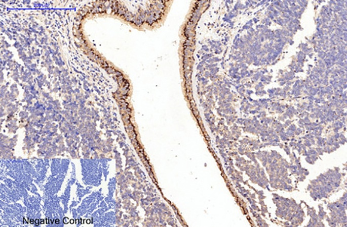
Immunohistochemistry (IHC) performance optimization and FFPE tissue validation make ABM40379 the go-to antibody for clinical and archival tissue analysis, the cornerstone of translational collagen research. FFPE tissues are the standard for long-term clinical sample storage and pathological analysis, but the formalin fixation process causes cross-linking of collagen proteins, masking epitopes and leading to weak or undetectable signal in many IHC assays. ABM40379 is specifically optimized for FFPE tissue detection, with a suggested starting dilution of 1:50–300 for IHC and experimental validation confirming 1:200 as the optimal working dilution for human lung cancer, rat kidney, and other FFPE tissue types. The antibody’s IHC performance is further enhanced by a validated antigen retrieval step—sodium citrate buffer (pH 6.0) heated to >98°C for 20 minutes— which effectively unmasks Type I Collagen epitopes without damaging tissue morphology or compromising antibody binding. Critically, ABM40379 produces minimal non-specific background staining in FFPE sections, with negative controls (secondary antibody only) showing no detectable signal— a hallmark of high antibody specificity that reduces experimental error and ensures the reliability of qualitative and semi-quantitative IHC data. This makes the antibody an ideal tool for clinical pathology research, where accurate Type I Collagen staining is essential for diagnosing collagen-related disorders and characterizing pathological tissue remodeling.
Immunofluorescence (IF) compatibility with a starting dilution of 1:50–200 expands ABM40379’s utility for spatial biology research, a fast-growing area of collagen science focused on the subcellular and tissue-level localization of Type I Collagen. Unlike IHC, IF enables the precise visualization of Type I Collagen’s spatial distribution relative to other cellular and matrix components, a key requirement for understanding its functional interactions in healthy and pathological tissues—for example, mapping Type I Collagen fiber networks in the tumor stroma and their association with cancer-associated fibroblasts, or tracking collagen deposition during embryonic connective tissue development and wound healing. ABM40379 delivers crisp, defined fluorescent signal for Type I Collagen in fresh/frozen tissues and cultured cells, avoiding the diffuse, non-specific staining that plagues low-quality collagen antibodies and enabling clear co-localization analysis with other molecular markers (e.g., fibroblast markers, bone morphogenetic proteins, tumor cell antigens). This IF performance aligns with the industry trend toward spatial omics and high-resolution tissue imaging, where the accurate visualization of matrix proteins is critical for decoding the complex architecture of connective tissues and their role in health and disease.
Optimized formulation and robust storage characteristics of ABM40379 ensure long-term antibody activity and experimental reproducibility, a critical consideration for research reagents that underpin consistent, publishable data. The antibody is supplied as a ready-to-use liquid solution at a concentration of 1 mg/ml, formulated in a buffered solution of PBS containing 50% glycerol, 0.5% BSA, and 0.02% sodium azide— a formulation engineered for maximum protein stability. Glycerol prevents ice crystal formation during freezing, a major cause of antibody denaturation; BSA acts as a protein stabilizer to maintain antibody structure and binding affinity; and sodium azide serves as a mild, broad-spectrum preservative safe for all in vitro IHC and IF applications. ABM40379 is stable for one year at -20°C from the date of shipment, with simple handling guidelines to preserve activity: aliquot the 30μl stock into small volumes (5–10μl) upon first use to avoid repeated freezing and thawing, centrifuge the vial after thawing to collect antibody adhered to the vial wall, and prepare fresh dilutions for each experiment. These features address a common industry challenge—reagent instability leading to inconsistent experimental results—and ensure that ABM40379 delivers reliable performance across months of research, even for labs with intermittent experimental workflows.
Exceptional cost-effectiveness and broad research applicability make ABM40379 an accessible and indispensable tool for labs of all sizes, from small academic research groups to large pharmaceutical and biotech companies focused on regenerative medicine and drug development. Priced at $109 for 30μl of 1 mg/ml antibody, ABM40379 offers unparalleled value: its high working dilutions (1:50–300 for IHC, 1:50–200 for IF) mean a single vial provides sufficient reagent for dozens of experiments, far outperforming more expensive Type I Collagen antibodies on the market. The antibody’s applications span a diverse range of high-impact research disciplines: skeletal biology (studying bone formation and osteogenesis imperfecta), dermatology (investigating skin aging and wound healing), oncology (profiling TME collagen remodeling), fibrosis research (characterizing liver, kidney, and lung fibrosis), and tissue engineering (validating collagen deposition in artificial scaffolds). This broad applicability aligns with the growing industry focus on collagen as a therapeutic target—for example, anti-fibrotic drugs that inhibit abnormal Type I Collagen deposition, or tissue engineering scaffolds that mimic native collagen structure for regenerative medicine. ABM40379’s ability to support research across all these areas makes it a cost-effective, multi-purpose reagent that drives progress in some of the most important areas of modern biology and medicine.
In conclusion, the Abbkine Collagen I Mouse Monoclonal Antibody (4H10) (ABM40379) emerges as the gold-standard tool for Type I Collagen research, combining uncompromising specificity, cross-species reactivity, validated performance across IHC and IF, and robust stability to address the core technical challenges of collagen detection (product link: https://www.abbkine.com/product/collagen-i-mouse-monoclonal-antibody-4h10-abm40379/). Its epitope-specific design eliminates cross-reactivity with other collagen subtypes, its human/mouse/rat validation streamlines translational research, its FFPE optimization enables reliable clinical tissue analysis, and its IF compatibility supports high-resolution spatial biology research—all while offering exceptional cost-effectiveness and long-term performance. For researchers investigating the structural and functional roles of Type I Collagen in health and disease, ABM40379 is more than a research reagent: it is a precision tool that unlocks new insights into collagen biology, accelerates translational research, and lays the groundwork for the development of novel diagnostics and therapeutics for collagen-related disorders and diseases. As the study of the extracellular matrix and connective tissue biology continues to expand, ABM40379 stands as a trusted and essential partner for researchers pushing the boundaries of this dynamic field.


