| Product name | GFP Monoclonal Antibody |
| Immunogen | Recombinant Protein |
| Host | Mouse |
| Applications | IP, WB |
| Applications notes | Optimal working dilutions should be determined experimentally by the investigator. Suggested starting dilutions are as follows: WB (1:5000), IP (1:200). |
| Clonality | Monoclonal |
| Preparation method | The antibody was affinity-purified from mouse ascites by affinity-chromatography using specific immunogen |
| Alternative | GFP; Green fluorescent protein |
| Formulation | Liquid solution |
| Concentration | 1 mg/ml |
| Storage buffer | PBS, pH 7.4, containing 0.02% Sodium Azide as preservative and 50% Glycerol. |
| Storage instructions | Stable for one year at -20°C from date of shipment. For maximum recovery of product, centrifuge the original vial after thawing and prior to removing the cap. Aliquot to avoid repeated freezing and thawing. |
| Shipping | Gel pack with blue ice. |
| Precautions | The product listed herein is for research use only and is not intended for use in human or clinical diagnosis. Suggested applications of our products are not recommendations to use our products in violation of any patent or as a license. We cannot be responsible for patent infringements or other violations that may occur with the use of this product. |
| Background | Green Fluorescent Protein (GFP) has quickly become a powerful research tool for assessing gene expression and subcellular protein distribution in fixed or living cells. GFP is excited by and brightly fluoresces when exposed to UV or blue light. This feature makes it ideal as a marker for use in fluorescence microscopy, cytometry, tagging fusion proteins, and assaying transcriptional regulation from gene promoters in vivo. Numerous GFP variants with enhanced and shifted emission spectra (blue, green, and yellow) have been developed through amino acid substitutions at specific residues. |
| Alternative | GFP; Green fluorescent protein |
| Others | The antibody detects RFP, YFP, CFP. |
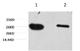
Fig.1. Western blot analysis of GFP transfected Hela, diluted at 1) 1:5000, 2) 1:10000.
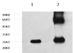
Fig.2. 1) control, 2) IP products, antibody dilution 1:200.
Author: Gu D, Wu S, Yu Z Publication name:Horticulture Research IF:7.291
Author:H Peng, L Jin, Q Zhang, Y Shen, Publication name:Traditional Chinese Medicine and Small Molecule Treatment of Tumors IF:2.65
You must be logged in to post a review.
1.The species of antibody reactivity should be the sample species that can be matched normally after Abbkine R&D experts have passed strict scientific verification. If your sample is not within the range of reactivity, in order to improve the efficiency and results of your experiment, it is not suggested to try other species. Otherwise, it may lead to sample mismatch and affect the effect of your experiment.
2.Please aliquot the antibody received as soon as possible and store it at -20℃, avoid repeated freezing and thawing, and use it within one year.
Welcome any form of communications, and better service will be provided here.
Tell: +1-404-854-0155
Email: service@abbkine.com
Support Email: support@abbkine.com
Address: 3052 Stroop Hill Road, Apt 203, Atlanta 30303, Georgia, United States of America


Reviews
There are no reviews yet.